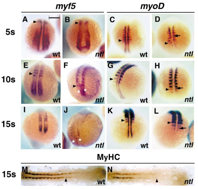
Fig. 5 Failure of adaxial myogenesis in posterior no tail embryos occurs at the Mrf induction stage. Analysis of 5 somite (A–D), 10 somite (E–H), and 15 somite (I–N) ntl embryos (B, D, F, H, J, L, N) reveals that adaxial myf5 (A, B, E, F, I, J) and myoD (C, D, G, H, K, L) expression is severely reduced at all stages and positions examined, compared to wild-type or heterozygous siblings (A, C, E, G, I, K, M). In contrast, lateral somitic expression levels of both myf5 and myoD appear unaffected by the ntl mutation at early stages. Note the delayed appearance of substantial medial myoD mRNA at somitic levels (arrows, D, H, L). myf5 mRNA accumulation in the tailbud tip is decreased at later stages of ntl development (asterisks, F) and is almost lost caudally in ntl embryos as outgrowth ceases (asterisks in J, dorsal three-quarter view, tailbud at bottom). Wholemount visualisation of all sarcomeric MyHC shows that adaxial muscle differentiation is substantially delayed in the 15 somite stage ntl embryo (M, N dorsal view, anterior to left). Bar, 200 μm in A–L and 164 μm in M, N.
Reprinted from Developmental Biology, 236(1), Coutelle, O., Blagden, C.S., Hampson, R., Halai, C., Rigby, P.W.J., and Hughes, S.M., Hedgehog signalling is required for maintenance of myf5 and myoD expression and timely terminal differentiation in zebrafish adaxial myogenesis, 136-150, Copyright (2001) with permission from Elsevier. Full text @ Dev. Biol.

