
|
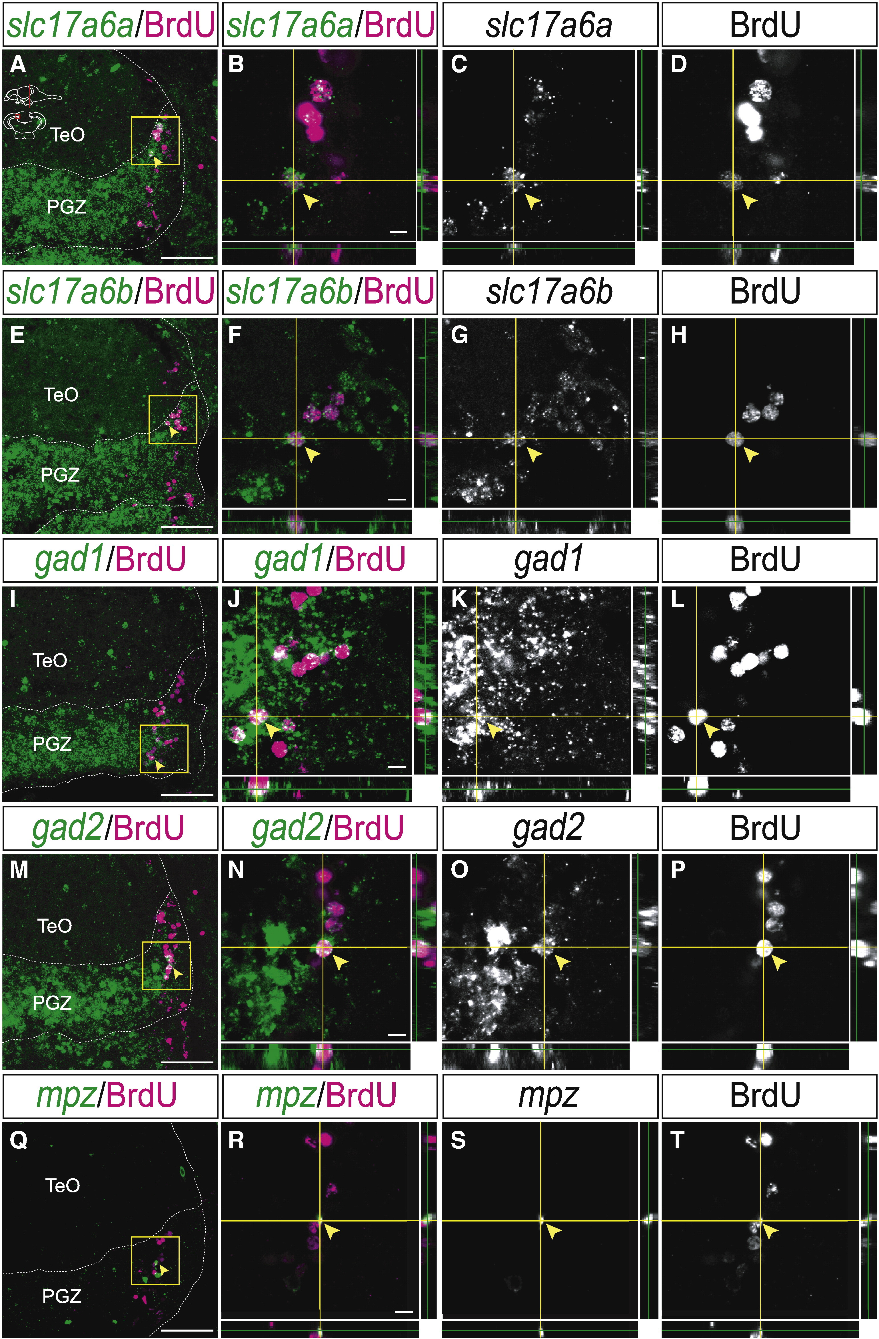
Fig. 7 Neural stem/progenitor cells in the mitotic region of the PGZ differentiate into the glutamatergic or GABAergic neurons and oligodendrocytes. (A?P) Expression of glutamatergic and GABAergic neuronal markers in the BrdU-positive cells in the PGZ of the adult zebrafish optic tectum at 1 month post-BrdU administration (14 μm transverse sections, stacked images, dorsal top). (A?H) Some of the BrdU-positive cells express glutamatergic neuronal markers, slc17a6a (A?D, arrowheads) and slc17a6b (E?H, arrowheads) (B?D: high magnification of yellow-boxed area in A, F?H: high magnification of yellow-boxed area in E). (I?P) Some of the BrdU-positive cells express GABAergic neuronal markers, gad1 (I?L, arrowheads) and gad2 (M?P, arrowheads) (J?L: high magnification of yellow-boxed area in I, N?P: high magnification of yellow-boxed area in M). (Q?T) Expression of oligodendrocyte marker, mpz in the BrdU-positive cells in the PGZ of the adult zebrafish optic tectum at 1 month post-BrdU administration (arrowheads) (14 μm transverse sections, stacked images, dorsal top) (R?T: high magnification of the yellow-boxed area in Q). CCe, corpus cerebelli; PGZ, periventricular gray zone; TeO, tectum opticum. Scale bars: 50 μm in A, E, I, M, Q; 5 μm in B, F, J, N, R.
Reprinted from Developmental Biology, 342(1), Ito, Y., Tanaka, H., Okamoto, H., and Ohshima, T., Characterization of neural stem cells and their progeny in the adult zebrafish optic tectum, 26-38, Copyright (2010) with permission from Elsevier. Full text @ Dev. Biol.