IMAGE
Fig. 8
Image
Figure Caption
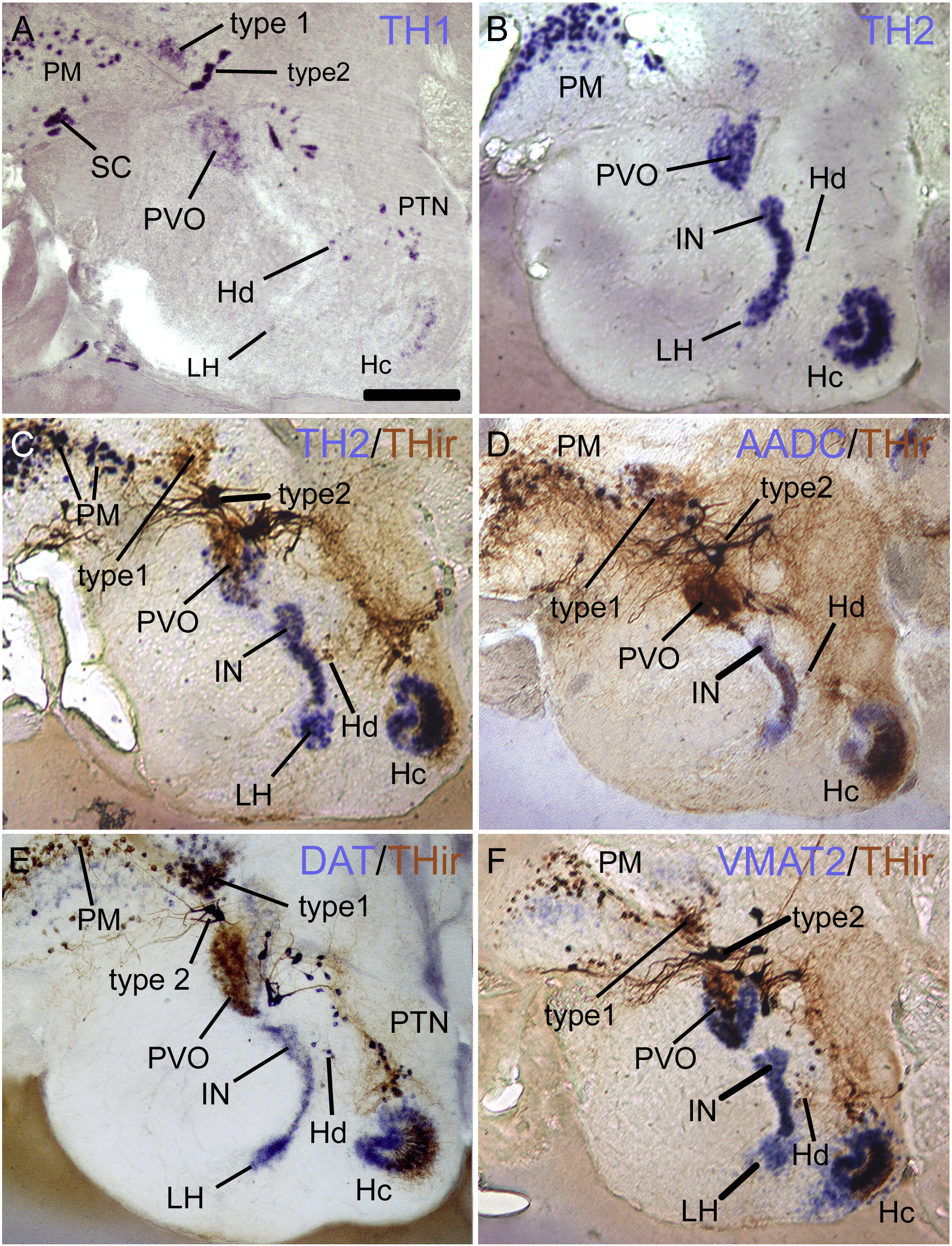
Fig. 8 Sagittal sections through preoptic and adjacent diencephalic regions, showing TH1 single ISH (A), TH2 single ISH (B), and double-labeling for TH2 ISH (C), AADC ISH (D), DAT ISH (E), or VMAT2 ISH (F) with THir, respectively. The localization of TH1 transcript is practically identical to THir. Note that there are many double-labeled (dark brown) cells in the preoptic area (PM is shown mostly), while many TH2, AADC, DAT, and VMAT2 cells without THir are found in the hypothalamus. Scale bar = 200 μm.
Figure Data
Acknowledgments
This image is the copyrighted work of the attributed author or publisher, and
ZFIN has permission only to display this image to its users.
Additional permissions should be obtained from the applicable author or publisher of the image.
Reprinted from Molecular and cellular neurosciences, 43(4), Yamamoto, K., Ruuskanen, J.O., Wullimann, M.F., and Vernier, P., Two tyrosine hydroxylase genes in vertebrates: New dopaminergic territories revealed in the zebrafish brain, 394-402, Copyright (2010) with permission from Elsevier. Full text @ Mol. Cell Neurosci.

