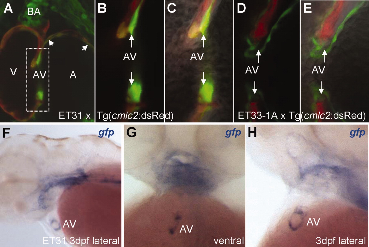
Fig. 4 The CET line, ET31, marks a unique subset of myocardium at the early A-V valve. A: ET31 crossed with Tg(cmlc2:dsRed) showing a subset of myocardium positive for EGFP at the A-V junction and chambers (arrowhead). Note the EGFP expression at the BA. B, C: High magnification of A-V junction reveals differential GFP intensity between a different subset of myocardial cells. Arrows point to non-overlapped ET31 EGFP expression. D, E: High magnification of A-V junction lined with endocardial cells adjacent to cmlc2 marked myocardium (shown for comparison). Arrows point to A-V junction that ET31 marks. F-H: anti-gfp WISH demonstrates that the expression domain at the A-V node represents a ring. A-C: Double transgenic embryos of ET31 and Tg(cmlc2:dsRed). D, E, Double transgenic embryos of ET33-1A and Tg(cmlc2:dsRed). B, D: Fluorescent images. C, E: Composite fluorescent/DIC images to reveal cellular morphology. A, atrium; BA, bulbus arteriosus; A-V, atrio-ventricular valve; V, ventricle.
Image
Figure Caption
Figure Data
Acknowledgments
This image is the copyrighted work of the attributed author or publisher, and
ZFIN has permission only to display this image to its users.
Additional permissions should be obtained from the applicable author or publisher of the image.
Full text @ Dev. Dyn.

