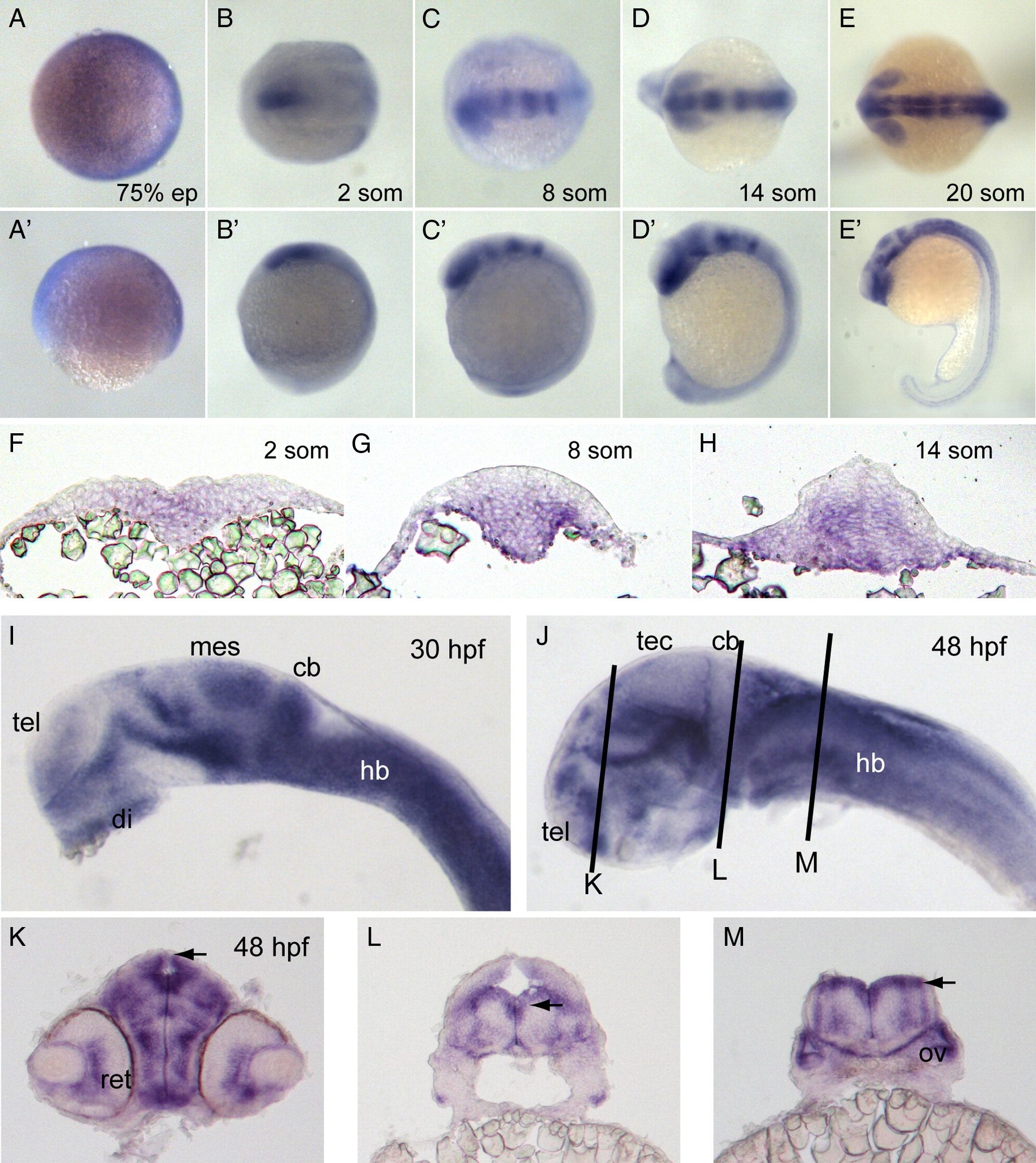
Fig. 3 Expression of pcdh19. (A?E) In situ hybridization of embryos labeled with pcdh19 riboprobe against cadherin repeats 1?3 at 75% epiboly (A), 2 somites (B), 8 somites (C), 14 somites (D) and 20 somites (E). Dorsal views are shown in A?E and lateral views are shown in A′?E′. Pcdh19 is expressed broadly and diffusely at 75% epiboly. At 2 somite stage, pcdh19 is expressed in the medial anterior neural plate. By 8 somite stage, pcdh19 expression exhibits a banding pattern, with the anterior band encompassing the forebrain and eye primordia and additional bands present in the midbrain and hindbrain. These bands become sharper by 14 somite stage. By 20 somite stage, the pattern in the brain has become more complex, with expression in the eye, hypothalamus, ventral midbrain and the tectum. In addition, expression has become stronger and more uniform throughout the hindbrain. In the trunk region, pcdh19 is expressed in cells along the midline. F?H, Cross-sections of embryos labeled with pcdh19 riboprobe at 2 somite stage (F), 8 somite stage (G) and 14 somite stage (H). At 2 somite stage, pcdh19 is expressed medially in cells throughout the dorsoventral extent of the neural plate. As development proceeds, pcdh19 expression is concentrated more ventrally in both the neural keel and neural rod. I?J, In situ hybridizations reveal more distinctive expression patterns at 30 hpf (I) and 48 hpf (J). Abbreviations of brain regions are as follows: tel, telencephalon; di, diencephalon; mes, mesencephalon; cb, cerebellum; hb, hindbrain; tec, optic tectum. K?M, Cross-sections of 48 hpf embryos at the three planes of section indicated in (J). Both the retina (ret) and the otic vesicle (ov) are labeled with the pcdh19 riboprobe. Notably, the neuroepithelia that line the ventricles (arrowheads) express high levels of pcdh19.
Reprinted from Developmental Biology, 334(1), Emond, M.R., Biswas, S., and Jontes, J.D., Protocadherin-19 is essential for early steps in brain morphogenesis, 72-83, Copyright (2009) with permission from Elsevier. Full text @ Dev. Biol.

