Fig. 2
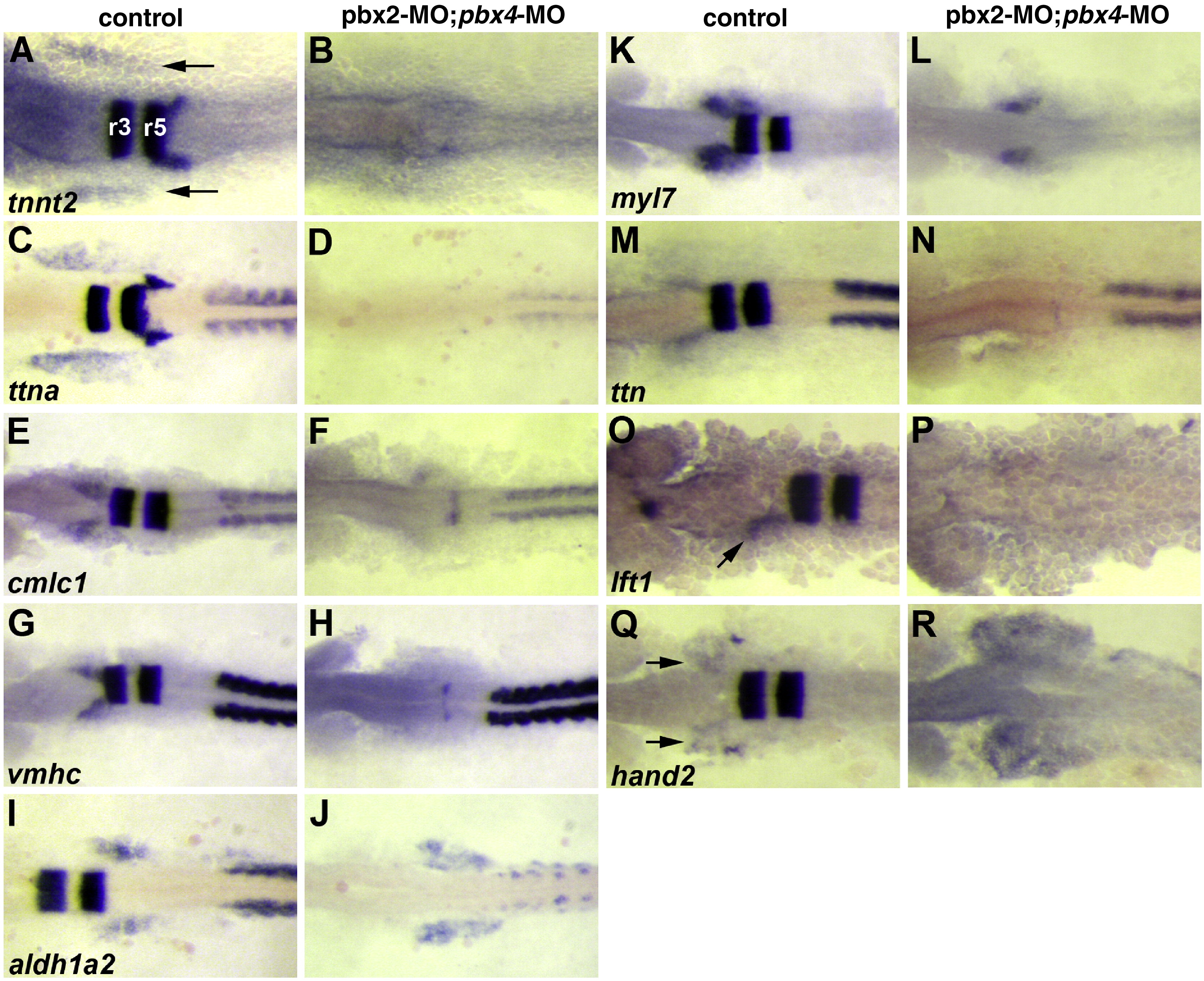
Fig. 2 Validation of microarray-identified Pbx-dependent heart genes. (A?R) RNA in situ expression of genes from Table 1 in (A, C, E, G, I, K, M, O, Q) wild-type control or (B, D, F, H, J, L, N, P, R) pbx2-MO; pbx4-MO embryos. (A?D) are at 10 somite stage (10s). (E?N,Q?R) are at 18s. (O?P) are at 20s to achieve more robust expression of lft1 in controls. krox-20, labeled in hindbrain rhombomeres r3 and r5 in (A), is included in all in situs to control for loss of Pbx. The wild-type heart primordium expression domains of tnnt2, ttna, cmlc1, vmhc, myl7, lft1 and hand2 are just anterior-lateral to r3 krox-20 expression (arrows in A, O, Q). n ≥ 10 for each marker in control or pbx2-MO; pbx4-MO. Embryos are shown in dorsal view, anterior towards the left.
Reprinted from Developmental Biology, 333(2), Maves, L., Tyler, A., Moens, C.B., and Tapscott, S.J., Pbx acts with Hand2 in early myocardial differentiation, 409-418, Copyright (2009) with permission from Elsevier. Full text @ Dev. Biol.

