
Image
|
Figure Caption
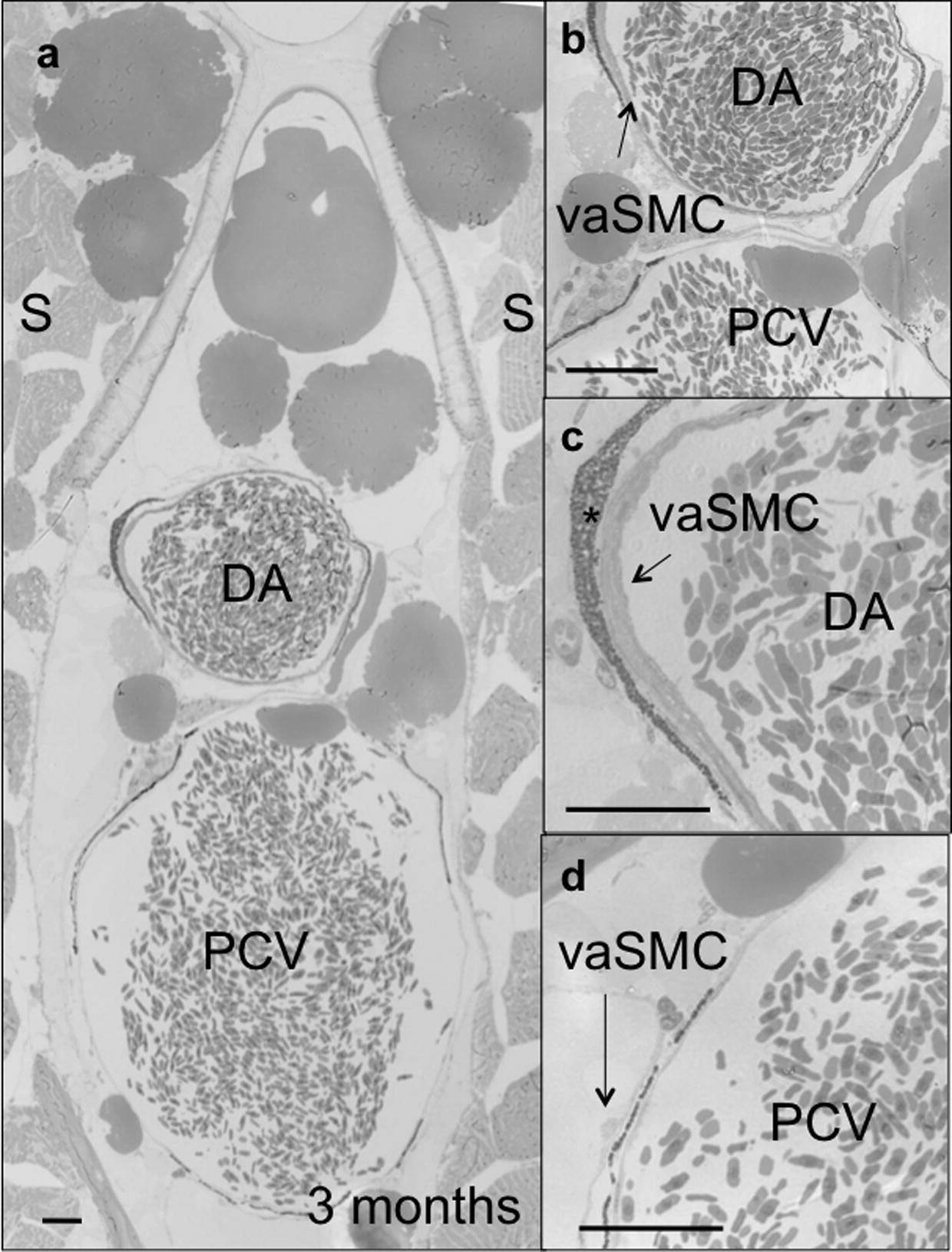
Fig. 1 Histological analyses of the dorsal aorta and posterior cardinal vein of 3 months old zebrafish. (a?d) Toluidine blue-stained transverse sections of the trunk vasculature showing the dorsal aorta (DA) and posterior cardinal vein (PCV). A thick layer (arrows in (b and c)) of vascular SMCs (vaSMCs) is present around the DA but not the PCV, where single vaSMCs (arrow in (d)) are interspersed along the circumference of the PCV. Scale bars, 100 μm. *Peri-aortic melanocytes. Sections are at the level of the 10th somite.
Acknowledgments
This image is the copyrighted work of the attributed author or publisher, and
ZFIN has permission only to display this image to its users.
Additional permissions should be obtained from the applicable author or publisher of the image.
Reprinted from Mechanisms of Development, 126(8-9), Santoro, M.M., Pesce, G., and Stainier, D.Y., Characterization of vascular mural cells during zebrafish development, 638-649, Copyright (2009) with permission from Elsevier. Full text @ Mech. Dev.