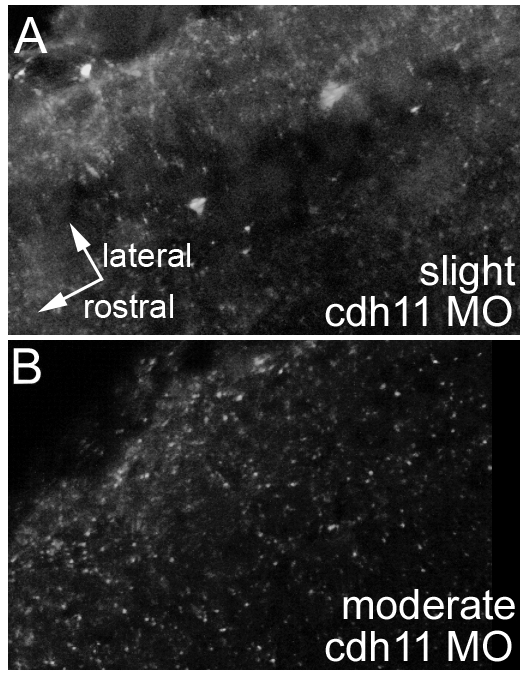
Fig. S2 Cdh11 morpholino-induced expression reduction through the entire otic vesicle. To determine whether optical section selection in Figure 1B,C minimized strongly labeled structures (like otoliths), we collected image volumes from the same zebrafish embryos labeled using Cdh11-specific antibodies shown in Figure 1B,C (i.e., injected with cdh11 splice-site blocking antisense morpholino oligonucleotide, as here in A and B). Instead of a projection of 5 optical sections as shown in Figure 1B,C, an image volume was collected through the entire otic vesicle of a slightly affected 30-hpf embryo (A; 45 optical sections, 0.3 μm z-step) and a moderately affected 30-hpf embryo (B; 25 optical sections, 0.3 μm z-step). Projection images were made in alpha blending mode using Voxx software. Adobe Photoshop 6 was used to prepare the figure. The otic vesicle was smaller in the moderately affected embryo, but both images (A and B) are projections of image volumes through the entire inner ear region of the embryos. A and B have identical orientation, with rostral and lateral as shown in A. These image volumes illustrate that only small amounts of Cdh11 labeling were detected in slightly affected phenotype embryos (A), and little or no Cdh11 labeling was detected above background in moderately affected (in B, background from 25 optical sections is combined) and severely affected (not shown) embryos. Laser power, gain settings, filters, and image processing were identical for A and B. Two-photon imaging was performed using Zeiss LSM510 Meta NLO, C-Apochromatic 40xW 1.2NA objective, TRITC-labeled secondary antibody, 800-nm excitation, and mounted in PBS.
Image
Figure Caption
Acknowledgments
This image is the copyrighted work of the attributed author or publisher, and
ZFIN has permission only to display this image to its users.
Additional permissions should be obtained from the applicable author or publisher of the image.
Full text @ Dev. Dyn.

