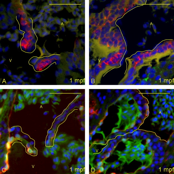
Fig. 6 Larger larvae have more pronounced maturation of the valves at 28 days postfertilization (dpf). A-D: Five-micrometer paraffin sections of flk1:GFP (GFL, green fluorescent protein) transgene-carrying zebrafish larvae were imaged to identify the structural appearance of the atrioventricular (AV) boundary and intra-luminal structure. Atria (A) and ventricles (V) are labeled (yellow letters), and yellow lines outline the extent of the AV structure (cushion and/or valve). A,B: Sections were immunostained with DAPI (4′,6-diamidine-2-phenylidole-dihydrochloride; blue), anti-Versican (red) and anti-Collagen I (green). Whereas the smaller larva (A) has started deposition of Versican in the leaflet, the larger larva (B) has thickened considerably more, with increased deposition of Versican and Collagen I. C,D: From separate larvae than shown in A,B, sections were stained with DAPI (blue), anti-GFP (red), and anti-Collagen II (green). Again, the larger larva (D) has significantly more deposition of collagen than the smaller larva (C), despite being at the same age. Scale bar = 20 μm.
Image
Figure Caption
Acknowledgments
This image is the copyrighted work of the attributed author or publisher, and
ZFIN has permission only to display this image to its users.
Additional permissions should be obtained from the applicable author or publisher of the image.
Full text @ Dev. Dyn.

