
|
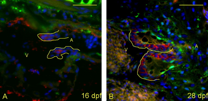
Fig. 4 Maturation of the valve leaflets initiates between 16 and 28 days postfertilization (dpf). A,B: Five-micrometer paraffin sections of zebrafish larvae were imaged to identify the structural appearance of the AV boundary and intraluminal structure. Atria (A) and ventricles (V) are labeled (yellow letters), and yellow lines outline the extent of the atrioventricular (AV) structure (cushion and/or valve). Cells were stained with DAPI (4′,6-diamidine-2-phenylidole-dihydrochloride; blue), anti-collagen II (green) and anti-versican (red). A: At 16 dpf, the valve leaflets are highly cellular, with little collagen or versican present. B: By 28 dpf, the leaflets start to thicken with deposition of significant collagen fibers and versican.