
|
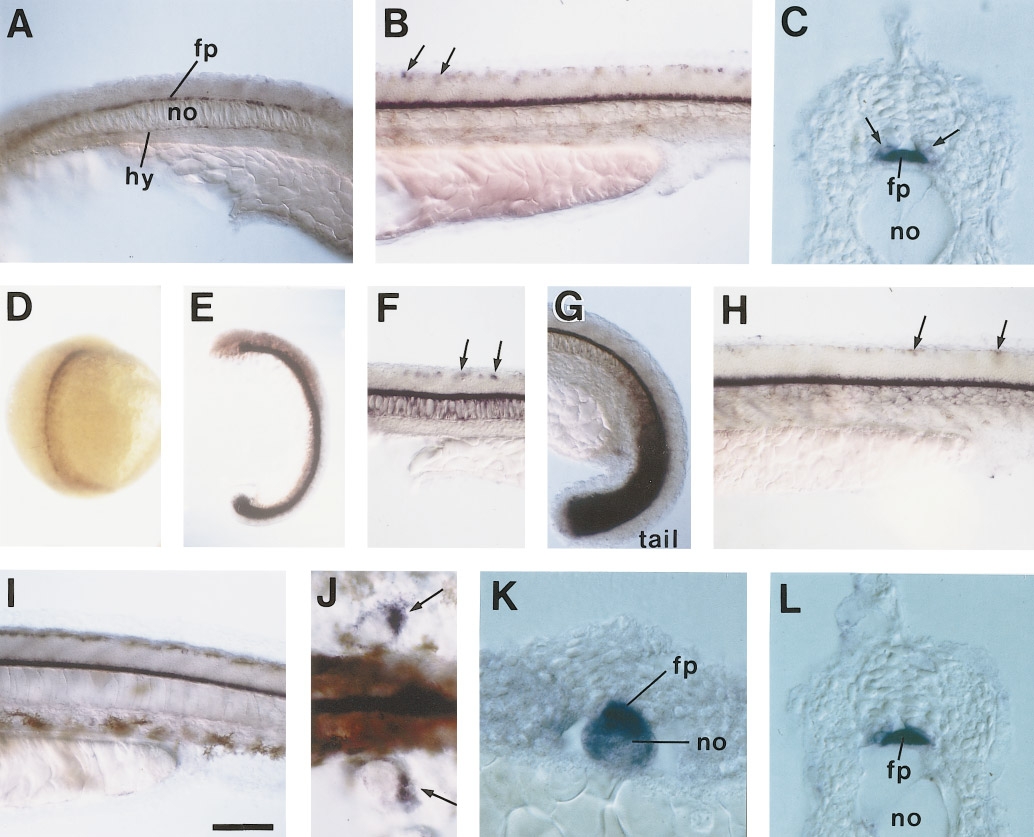
Fig. 5 (A?C) F-spondin1 and (D?L) F-spondin2 mRNA expression in developing embryos. (A) Lateral view of a 20-h embryo. F-spondin1 is clearly expressed in the floor plate and weakly expressed in the hypochord. (B) Lateral view of a 28-h embryo. F-spondin1 is expressed around the floor plate. In addition, several dorsal cells in the spinal cord express F-spondin1 (arrows). (C) Cross section of a 28-h embryo. F-spondin1 is strongly expressed in the floor plate. In addition, F-spondin1 appears to be weakly expressed in the cells adjacent to the floor plate (arrows). (D) F-spondin2 expression in a 13-h embryo. Dorsal view. F-spondin2 is expressed around the axial mesoderm and weakly in the somites. (E) Lateral view of a 17-h embryo. (F and G) Lateral view of 19-h embryos in the trunk (F) and the tail (G). In addition to the expression around the floor plate, F-spondin2 is also weakly expressed in the notochord and strongly expressed in the tail bud. (H and I) Lateral view of 28- (H) and 36-h (I) embryos. Arrows in (F) and (H) indicate F-spondin2-positive cells in the dorsal spinal cord. (J) Dorsal view of a 36-h embryo in the pectoral fin bud region. F-spondin2 is expressed in the posterior region of the pectoral finbuds (arrows) (K) Cross section of a 15-h embryo. (L) Cross section of a 28-h embryo. fp, floor plate; no, notochord; hy, hypochord. Scale bar, 100 μm in A, B, and F?J; 17 μm in C, K, and L; 250 μm in D; and 160 μm in E.
Reprinted from Developmental Biology, 192, Higashijima, S., Nose, A., Eguchi, G., Hotta, Y., and Okamoto, H., Mindin/F-spondin family: novel ECM proteins expressed in the zebrafish embryonic axis, 211-227, Copyright (1997) with permission from Elsevier. Full text @ Dev. Biol.