
|
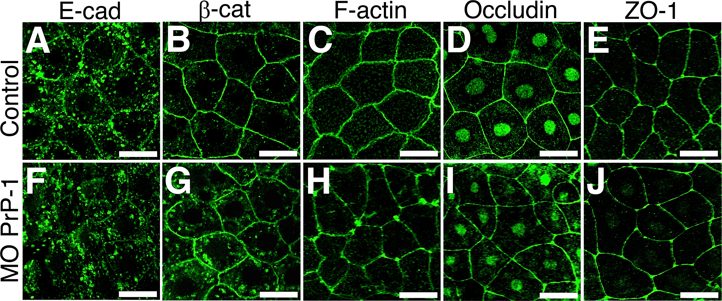
Fig. 8 PrP-1 Regulation of Adherens, but Not Tight Junctions, in EVL Cells
Differences in the subcellular distribution of various cell junction components between control and PrP-1 morphant embryos (MO) were evaluated in the polarized epithelial cells of the EVL at the shield stage (6 hpf). In control embryos, a marked membrane localization pattern can be seen for adherens junction (E-cadherin [E-cad], β-catenin [β-cat], and F-actin) (A?C) and tight junction markers (Occludin and ZO-1) (D and E). PrP-1 knockdown induces partial mislocalization of adherens junction components (F?H) but does not affect the distribution of classical tight junction markers (I and J). Scale bars indicate 20 μm.