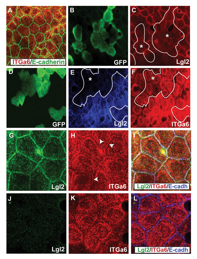
Fig. S1 Early localisation of Itga6 in lgl2 mutants and lgl2 morphant clones. (A-F) Co-immunostaining using anti-Itga6 (red) and anti-E-cadherin (A) antibodies; anti-GFP (B) and anti-Lgl2 (C) antibodies; anti-GFP (D), anti-Lgl2 (E) and anti-Itga6 (F) antibodies. The localisation of Itga6 is not altered in the basal epidermis of lgl2 larvae at 3 dpf (A). In the basal epidermal clones marked with GFP (B), lgl2 morpholino specifically knockdowns Lgl2 expression (C). In GFP-marked basal epidermal clones (D), wherein Lgl2 expression is attenuated (E), Itga6 localisation at the basal and lateral domains remains unperturbed. (G-L) Co-immunostainings using anti-Lgl2 (G,J), anti-Itga6 (H,K) and anti-E-cadherin (blue in the overlays; I,L) antibodies. In comparison to wild type (G-I), in the lgl2 mutant (J-L) at 3.75 dpf lateral Itga6 localisation is selectively altered. Asterisks, epidermal clones. Arrowheads in H mark the lateral Itga6 localisation in wild-type basal epidermis.
Image
Figure Caption
Acknowledgments
This image is the copyrighted work of the attributed author or publisher, and
ZFIN has permission only to display this image to its users.
Additional permissions should be obtained from the applicable author or publisher of the image.
Full text @ Development

