
|
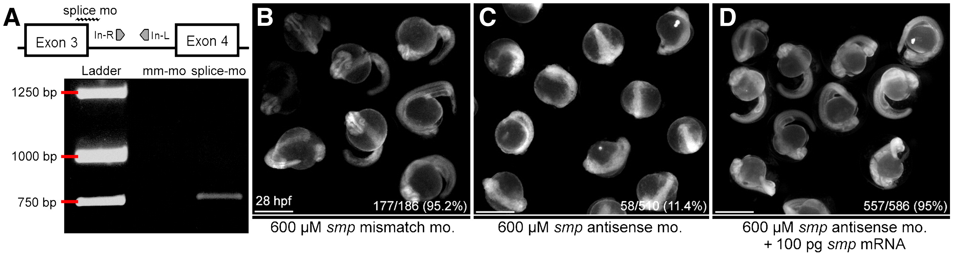
Fig. S2 Morpholino-mediated knock-down of smp is specific and can be rescued. (A) Diagram of the genomic region encompassing exons 3 and 4 as well as intron 3 of smp. Symbols mark positions of the antisense splice-junction morpholino (splice mo) and PCR primers (In-R and In-L). Underlying panel is a gel picture with DNA ladder and RT-PCR of mismatch-morpholino-transfected (mm-mo) and antisense-splice-junction-morpholino-transfected (splice mo) embryo extracts. (B) 28 hpf embryos after injection with mm-mo. (C) Embryos at the same stage after injection with splice-mo. (D) Rescued phenotype of same-staged embryos after co-injection of smp mRNA with splice-mo. Numbers indicate embryos showing wild-type phenotype/total injected embryos. The percents are the ratios of observed wild-type phenotype to total. Embryos in (B?D) show emissions from fluorescein-tagged morpholinos. Scale bars equal 500 μm.
Reprinted from Developmental Biology, 325(2), Kizil, C., Otto, G.W., Geisler, R., N�sslein-Volhard, C., and Antos, C.L., Simplet controls cell proliferation and gene transcription during zebrafish caudal fin regeneration, 329-340, Copyright (2009) with permission from Elsevier. Full text @ Dev. Biol.