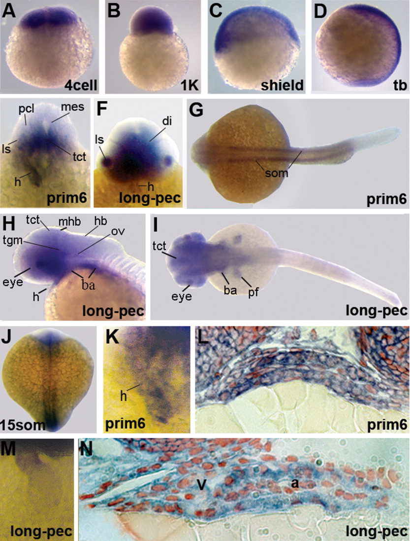
Fig. 2 Gene expression patterns of LMO7 during zebrafish development. Expression results of whole-mount in situ hybridization are shown for the seven stages indicated. A-D,G,H,J: In additional to embryos between cleavage and end of gastrulation (A-D), we present dorsal views of embryos at 15som, prim-6, and long-pec stage (G,H,J). E,F, I: A higher magnification of the dorsal view at prim-6 (F) and a lateral (I) and frontal (E) view of the head at long-pec stage are shown. K,M: Close-ups of the heart at prim-6 and long-pec stage are presented. L,N: Additionally, sections of the hearts at these stages are shown. ba, branchial arches; di, diencephalon; fp, floorplate; h, heart; hb, hindbrain; mb, midbrain; mhb, midbrain-hindbrain boundary; ls, lens; ov, otic vesicle; pf, pectoral fin buds; pcl, proliferative cell layer; tct, tectum; tegm, tegmentum.
Image
Figure Caption
Figure Data
Acknowledgments
This image is the copyrighted work of the attributed author or publisher, and
ZFIN has permission only to display this image to its users.
Additional permissions should be obtained from the applicable author or publisher of the image.
Full text @ Dev. Dyn.

