
|
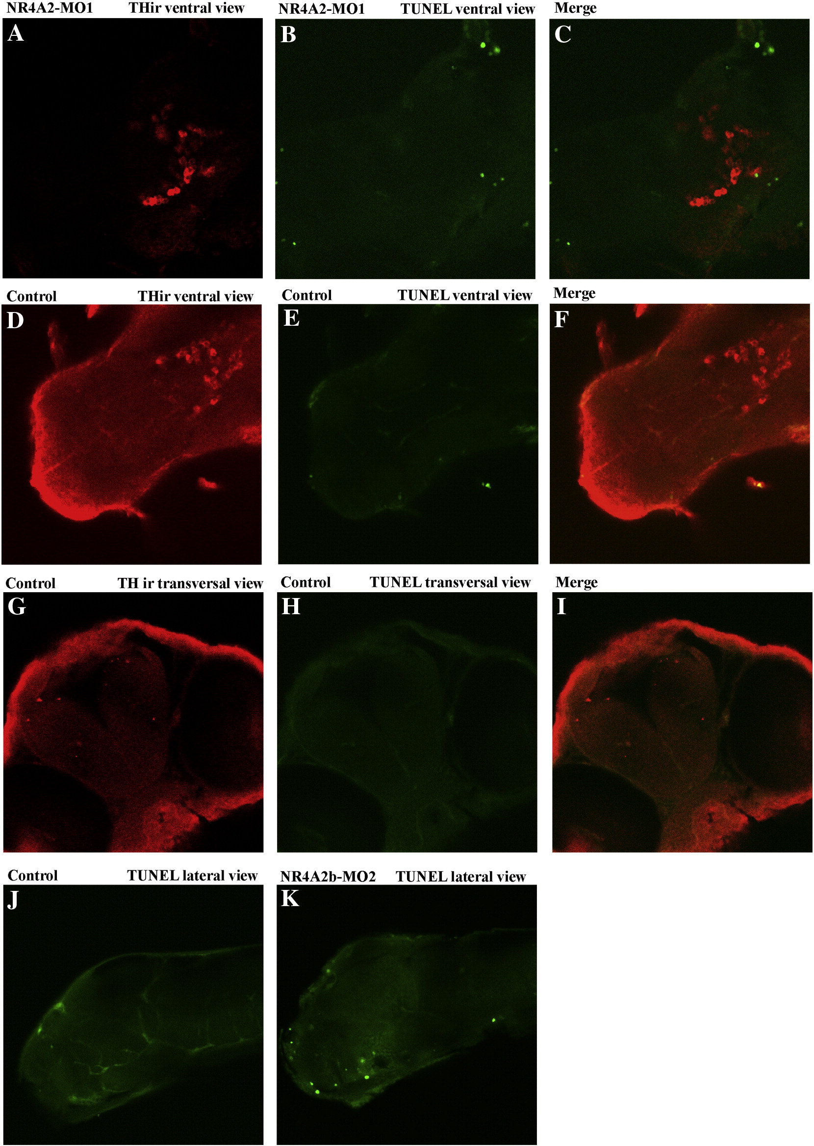
Fig. SD16 Compared apoptosis and TH expression in control and morphants at 72 hpf. (A–I) Double staining of THir (immunocytochemistry, red staining) and apoptotic cells (TUNEL, green staining), analyzed by scanning confocal microscopy. Sections of brain larvae at 72 hpf were obtained by confocal scanning. Merged pictures were made with by the image Image J software. (A,D,G) THir in red. (B,E,H) TUNEL staining in green. (C) Merged picture of hypothalamus of NR4A2-MO1 embryos. Merged picture of control hypothalamus (F) and telencephalon (I). TUNEL green staining in whole-mount 72 hpf control larvae(J) and NR4A2b-MO2 injected larvae (K).
Reprinted from Molecular and cellular neurosciences, 39(4), Blin, M., Norton, W., Bally-Cuif, L., and Vernier, P., NR4A2 controls the differentiation of selective dopaminergic nuclei in the zebrafish brain, 592-604, Copyright (2008) with permission from Elsevier. Full text @ Mol. Cell Neurosci.