
|
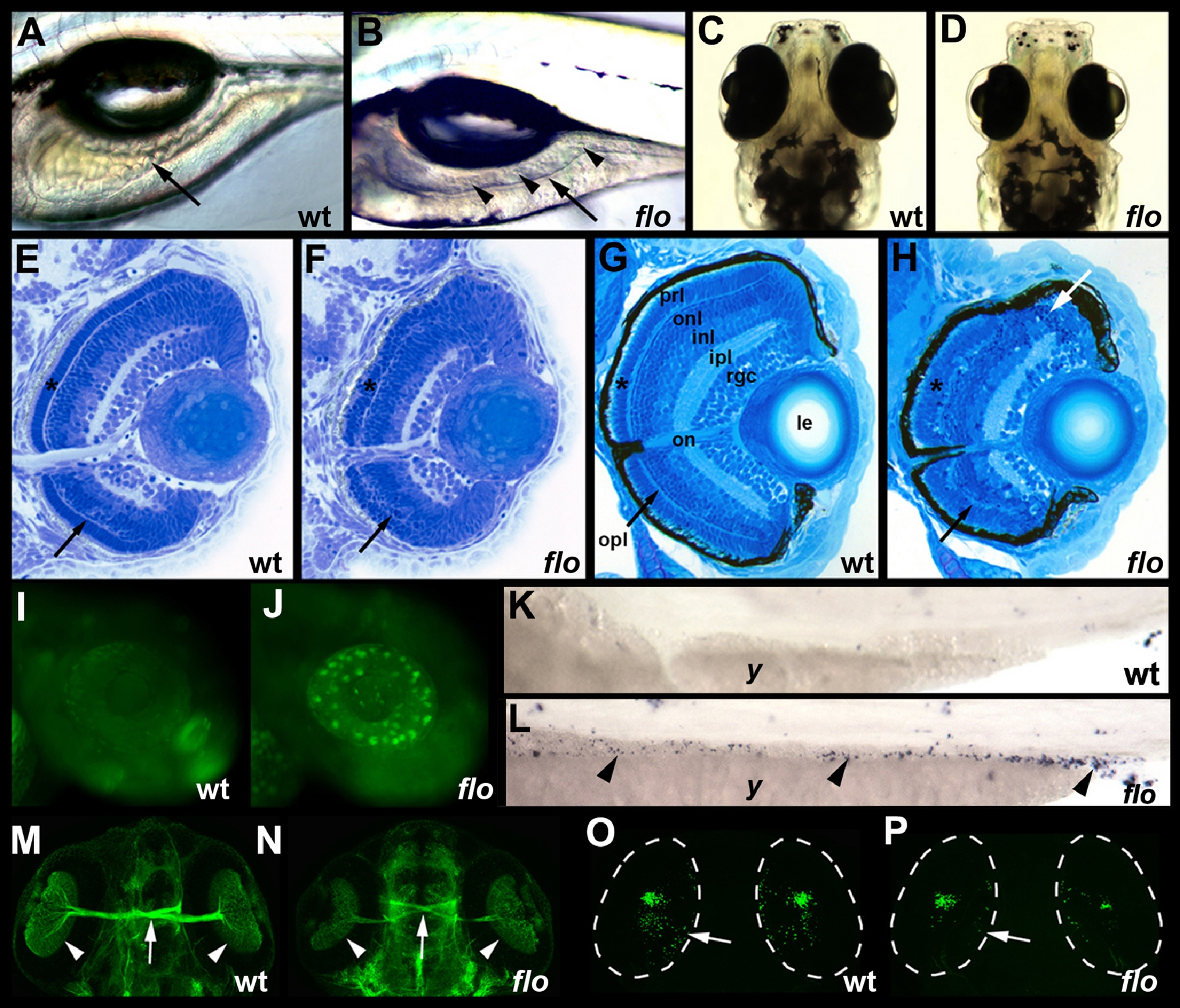
Fig. 1 flo intestinal and retinal defects.
(A,B) Lateral view of live 5 dpf wild type (wt) and flo larvae. The flo intestine lacks folds (arrow) and the lumen contains detached epithelial cells (arrowheads). (C,D) Dorsal view showing reduced size of the 5 dpf flo eye. (E,F) Histological cross section showing cells with condensed nuclei typical of apoptotic cells in the 60 hpf flo retina, and disorganization of the flo photoreceptor (*) and outer plexiform (arrow) layers. (G,H) Histological cross section showing marked disorganization of the 4 dpf flo retina and cells with condensed nuclei typical of apoptotic cells (white arrow). (I,J) Acridine orange staining showing apoptotic cells in the 48 hpf flo retina but not sibling wild types. (K,L) TUNEL staining showing apoptotic cells in the 75 hpf flo intestine (arrowheads) but not sibling wild types (anterior?left, posterior?right). (M,N) Dorsal view showing mild reduction in the number of flo retinal ganglion cells (arrowheads) and optic nerve diameter (arrow) identified with the Zn5 antibody. (O,P) Confocal projection of immunostained wt and flo larvae showing reduced rod cells in the flo retina including the large ventral cluster of cells and in the periphery of the mid retina (arrow). onl, outer nuclear layer; inl, inner nuclear layer; ipl, inner plexiform layer; rgc, retinal ganglion cell layer; on, optic nerve; le, lens; y, yolk.