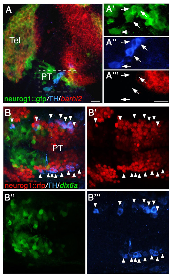
Fig. S2 All TH-positive DA neurons express neurog1 in the diencephalon. (A-A′″) Lateral view of neurog1::gfp transgenic reporter embryo (at 24 hpf), which was subjected to in situ hybridization with an antisense barhl2 probe followed by double immunofluorescence staining with antibodies against TH and GFP (A). High-magnification images of cells expressing neurog1, TH and barhl2 are shown in A′-A′″, respectively. Arrows indicate neurog1+ TH+ double-positive cells. (B-B′″) Dorsal view of double-transgenic neurog1::nRFP; dlx6a::GFP embryo (at 48 hpf), which was subjected to immunofluorescence staining with antibodies against TH (B). Arrowheads indicate neurog1+ TH+ double-positive neurons. Single-channel fluorescent images are presented in B′-B′″. PT, posterior tuberculum; RFP, red fluorescent protein; Tel, telencephalon. Scale bars: 20 μm.
Image
Figure Caption
Figure Data
Acknowledgments
This image is the copyrighted work of the attributed author or publisher, and
ZFIN has permission only to display this image to its users.
Additional permissions should be obtained from the applicable author or publisher of the image.
Full text @ Development

