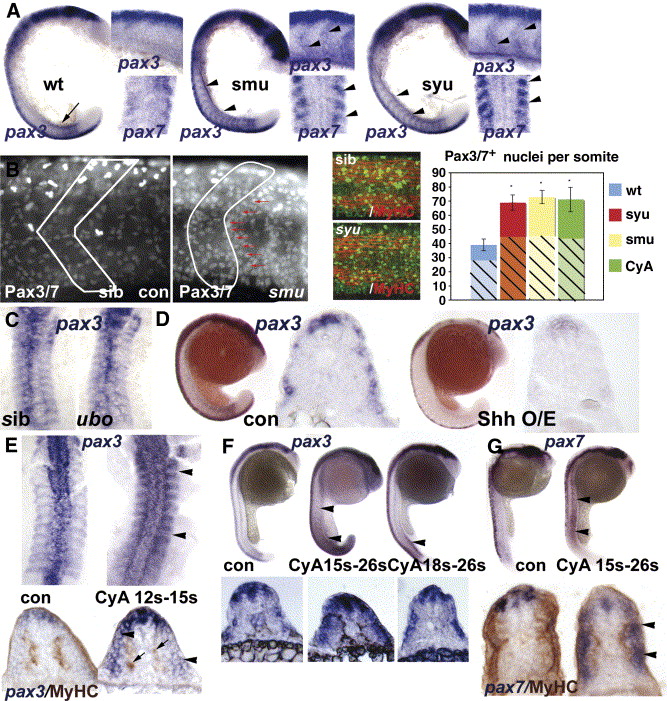
Fig. 4 Hedgehog signalling positively regulates somitic pax3/7 gene expression. (A) Flatmounts of wholemount in situ mRNA hybridisation for pax3 (at 18 s main panel, dorsal to left; upper inset, dorsal to top) and pax7 (at 15 s lower inset dorsal view of somites 3?6) in wild type, smu and syu mutant embryos. Note the transient expression of high level pax3 in nascent somites in wild type (arrow), and its persistence in anterior somites of mutants, particularly at the somite borders (arrowheads). (B) Confocal immunodetection of Pax3/7 protein increase in somites of 24 hpf smu (left panels, somite border outlined) and syu (central panels) embryos. Note the clustering of Pax-expressing cells near the anterior somite border (red arrows). Graph shows quantification of number of nuclei labelled with Pax3/7 (plain strong colour) or Pax7 (hatched pastel shade) in each condition from somites 12?16. Note that the Pax7 antibody gives less signal and more background, which probably explains the lower counts. Counts of three somites in 4?7 animals, error bars show standard deviation. Asterisks: P < 0.001. (C) Pax3 expression is indistinguishable in dorsal flatmounts of 15 s u-boot and sibling embryos. (D) Sonic hedgehog mRNA injection into the early embryo decreases pax3 mRNA at 18 s. Left panels wholemount, dorsal to left. Right panels, wholemount cryosection at yolk extension level. (E-G) Transient exposure to cyclopamine at the indicated stages leads to localised up-regulation of somitic pax3 (E, F) and pax7 (G). Upper panels wholemounts, dorsal to left. Lower panels, wholemount cryosection, dorsal to top. (E) Dorsal flatmount showing that treatment from 12 s to 15 s up-regulates pax3 in all somites (arrowheads) without affecting slow muscle formation (arrows, lower panels). (F) Treatment from 15 s up-regulates pax3 along most of rostrocaudal axis at 26 s (arrowheads), whereas treatment from 18 s has little effect in anterior somites, shown in transverse section below. (G) Cyclopamine from 15 s up-regulates pax7 throughout the axis at 26 s, but only in somitic tissue superficial to the slow muscle (arrowheads).
Reprinted from Developmental Biology, 302(2), Hammond, C.L., Hinits, Y., Osborn, D.P., Minchin, J.E., Tettamanti, G., and Hughes, S.M., Signals and myogenic regulatory factors restrict pax3 and pax7 expression to dermomyotome-like tissue in zebrafish, 504-521, Copyright (2007) with permission from Elsevier. Full text @ Dev. Biol.

