
Image
|
Figure Caption
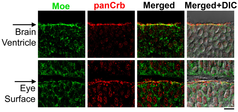
Fig. S2 Co-localization of Moe and panCrb in the brain and eye at 30 hours. Moe is shown in green (guinea pig anti-Moe) and panCrb in red (rabbit anti-CRB3). Images are merged with corresponding DIC images. Apical surfaces are indicated with arrows. Scale bar: 10 μm.
Figure Data
Acknowledgments
This image is the copyrighted work of the attributed author or publisher, and
ZFIN has permission only to display this image to its users.
Additional permissions should be obtained from the applicable author or publisher of the image.
Full text @ Development