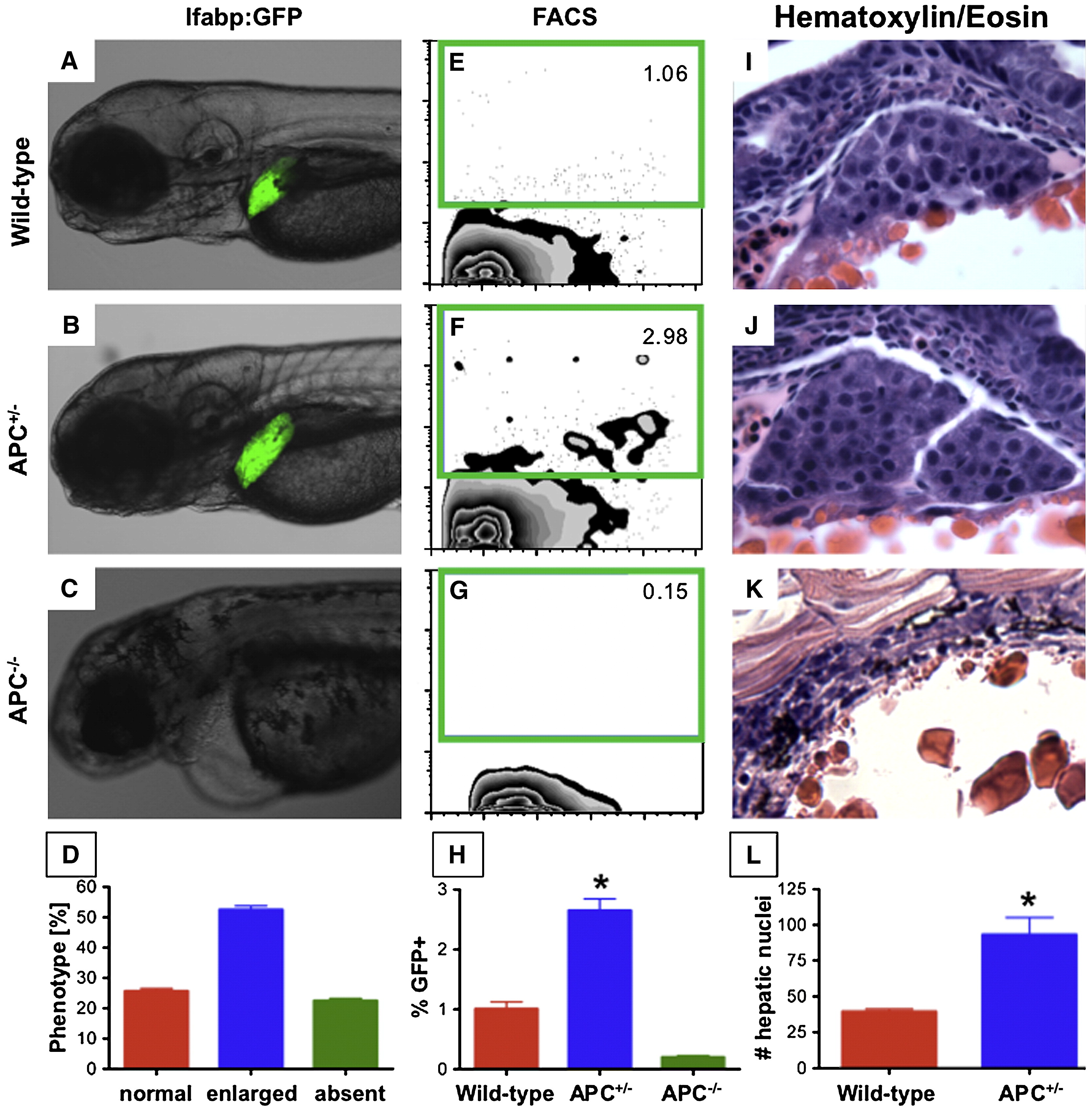
Fig. 1 APC loss has differential effects on liver development. Zebrafish embryos were analyzed at 72 hpf. (A?C) Fluorescence microscopy of progeny of an APC+/-; lfabp:GFP incross revealed differences in liver size. (D) Graphic representation of the liver phenotypes (5 independent clutches; n = 584) shows a Mendelian distribution. (E?G) Total hepatocytes per embryo were quantified by flow cytometry for GFP in each APC genotype (green gate) and confirmed the differential effects of APC loss on liver cell number (APC+/+ 1.00 ± 0.37% (% GFP+ hepatocytes of 20,000 total embryo cells counted ± SD); APC+/- 2.65 ± 0.62%; APC-/- 0.20 ± 0.10%; ANOVA, n = 10, p < 0.00001); (H) APC+/- embryos have significantly more hepatocytes than wild-type controls, while APC-/- embryos have no GFP+ hepatocytes. (I-K) H+E liver sections (10 μm) from wild-type, APC+/-, and APC-/- embryos (40x) corroborated the effects of APC mutations; (L) APC+/- embryos had increased hepatocytes per section compared to controls (93.3 ± 20.4 vs. 39.7 ± 3.1; t-test, n = 5, p = 0.004).
Reprinted from Developmental Biology, 320(1), Goessling, W., North, T.E., Lord, A.M., Ceol, C., Lee, S., Weidinger, G., Bourque, C., Strijbosch, R., Haramis, A.P., Puder, M., Clevers, H., Moon, R.T., and Zon, L.I., APC mutant zebrafish uncover a changing temporal requirement for wnt signaling in liver development, 161-174, Copyright (2008) with permission from Elsevier. Full text @ Dev. Biol.

