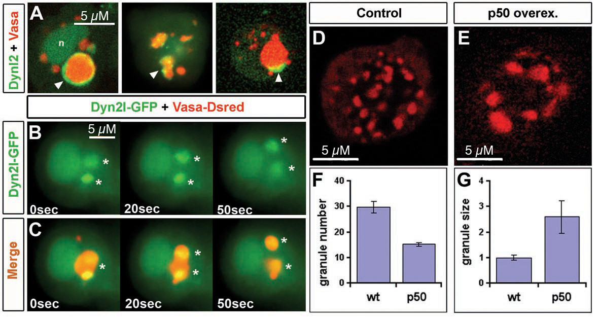
Fig. 6 Dynein localize to germ cell granules and is involved in granule fragmentation. A) DynL2-GFP is localized to nucleus, cytoplasm and to germ cell granules. Granule DynL2-GFP localization patterns are diverse, ranging from ring-like structures around large granules (left panel, arrowhead, nucleus is depicted by n), dots within the granules (arrowhead, middle panel) or accumulations can also be observed on the periphery of such structures (arrowhead, right panel). Interphase cells where analyzed in 6 to 10 hpf embryos. Note that Vasa-dsRed and DynL2-GFP colocalization preferentially occurs in the largest granule of the cell. B) Germ cells of 9 hpf embryos expressing DynL2-GFP were analyzed in vivo by epifluorescence microscopy. DynL2-GFP is localized to a large germ cell granule where two centers of high concentration are observed preceding the division of the granule (asterisks in B,C). C) Merge of B together with Vasa-dsRed to visualize the granules. D) Immunostaining of control germ cells of 24 hpf embryos showing the normal number (shown graphically in F) and size (shown graphically in G) of Vasa expressing granules n = 33 cells and 191 granules (G). Dynein inhibition in germ cells of 24 hpf embryos by p50 overexpression reduces the number of granules per germ cell while the size of germ cell granules increases. n = 116 cells and 154 granules (E,G). TTtest for F: p < 1E-8. TTest for G: p < 1E-14.
Image
Figure Caption
Figure Data
Acknowledgments
This image is the copyrighted work of the attributed author or publisher, and
ZFIN has permission only to display this image to its users.
Additional permissions should be obtained from the applicable author or publisher of the image.
Full text @ BMC Dev. Biol.

