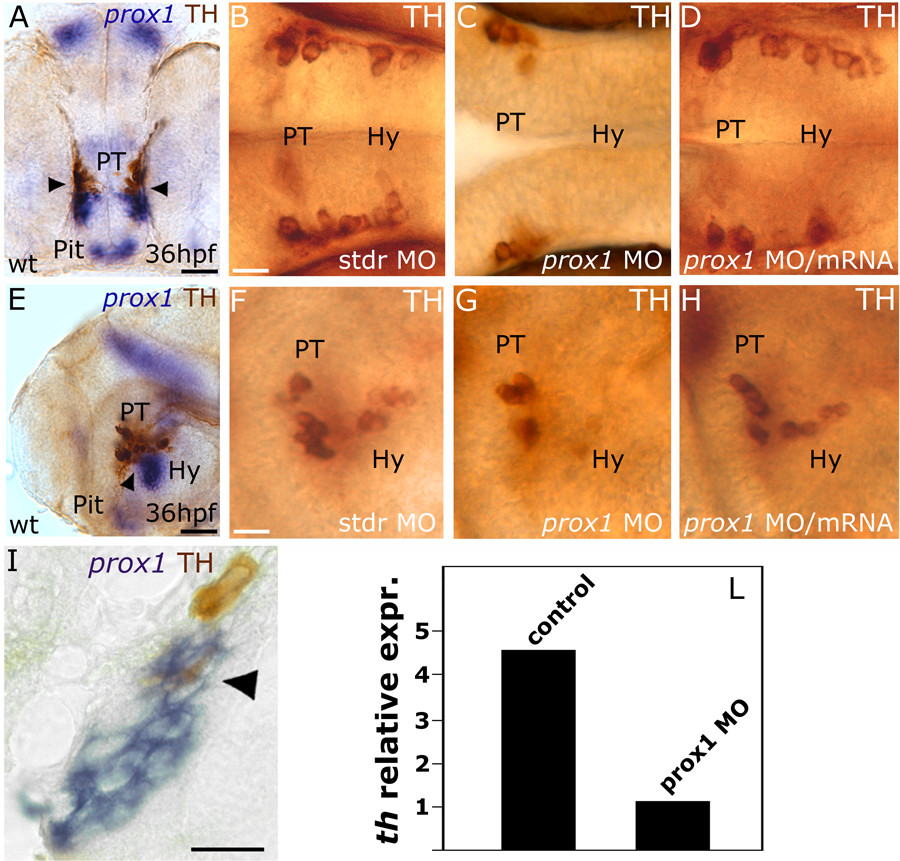
Fig. 2 prox1 is required for the development of CA neurons in the hypothalamus. Anterior is left in all panels except (A), frontal view. Dorsal is up, except for (B,C,D), dorsal view. Eyes or lens have been removed for better lateral viewing. (A,E,I) prox1 WISH combined with TH immunohistochemistry. Anti-TH antibody labels the PT and hypothalamic CA neurons at 36 hpf. Colabelling with prox1 is evident in a fraction of TH-positive neuroblasts in the hypothalamus (arrowheads), as also confirmed by the longitudinal section of the embryo (I). (C,G) microinjection of prox1 MO lowers the number of TH-labelled CA neurons in the hypothalamus in comparison to standard control injected embryos (B,F). (D,H) coinjection of prox1 mRNA and prox1 MO rescued the morphant phenotype. (L) Quantitative real time RT-PCR. TH-specific mRNA is almost five-fold decreased following prox1 MO injection. The result represents at least three independent experiments, and 18S was used as an internal control. The following abbreviations are used: posterior tuberculum (PT), pituitary (Pit), hypothalamus (Hy), standard control morpholino oligonucleotide (stdr MO). Scale bars indicate 10 μm (B,F,I) or 20 μm (A,E).
Image
Figure Caption
Figure Data
Acknowledgments
This image is the copyrighted work of the attributed author or publisher, and
ZFIN has permission only to display this image to its users.
Additional permissions should be obtained from the applicable author or publisher of the image.
Full text @ BMC Dev. Biol.

