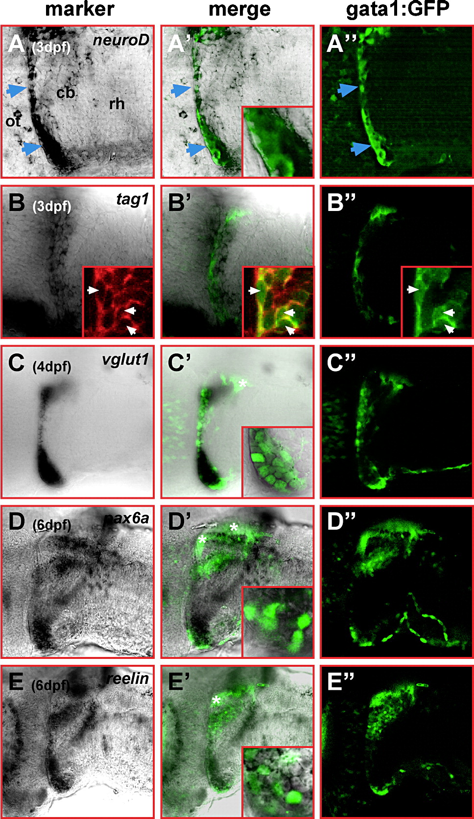
Fig. 2
Fig. 2 Cerebellar gata1:GFP cells show a granule cell expression profile. Single optical sections of lateral views of cerebella at 3 dpf (A?A″, B?B″), 4 dpf (C?C″) and dorsolateral views at 6 dpf (D?D″, E?E″), respectively, recorded by laser scanning confocal microscopy. In the left column, the cerebellar expression pattern of (A) neuroD, (B) tag1, (C) vglut1, (D) pax6a and (E) reelin is displayed. In the right column, cerebellar GFP-expression of the same optical section is displayed, while the overlap of both expression patterns is shown in the column in the middle with insets demonstrating the co-expression of GFP and the respective marker gene. Insets in panels B?B″ display Tag-1 expression detected by immunohistochemistry (B) co-localizing to GFP-expressing gata1:GFP cells (B′, B″ white arrowheads). GFP-expression in the dorsal-most cerebellum (white asterisks) is confined to parallel fibers (see Fig. 6), and thus, shows no co-localization with the analyzed marker gene expression. Abbr.: cb, cerebellum; ot, optic tectum; rh, rhombencephalon.
Reprinted from Developmental Biology, 313(1), Volkmann, K., Rieger, S., Babaryka, A., and Köster, R.W., The zebrafish cerebellar rhombic lip is spatially patterned in producing granule cell populations of different functional compartments, 167-180, Copyright (2008) with permission from Elsevier. Full text @ Dev. Biol.

