Fig. 3
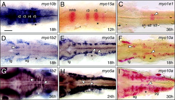
Fig. 3 Myosins exhibiting restricted expression patterns in the hindbrain. All panels show dorsal views of the hindbrain with anterior to the left. Asterisks mark the otic vesicle in each panel. (A and B) myo10b is expressed at varying levels in rhombomeres 2?5 (A), and myo15a is expressed in r3 and r5 (B), with both expressed at the midbrain?hindbrain boundary (mhb). (C) myo1e1 is weakly but specifically expressed in a mesendodermal domain spanning the 2nd and 3rd somites (arrowhead). (D and G) At 18 hpf, myo1b2 is expressed in sensory ganglia (tg, ag) and a cluster of cells in r2 (arrowhead, D). By 36 hpf (G), there is prominent expression in cells (arrowheads) at the boundaries of r5 and r6. (E and H) At 18 hpf, myo5a is expressed in sensory ganglia, and in differentiating neurons (arrowheads, E) within every rhombomere. By 24 hpf (H), expressing cells form a continuous column along the neural tube (arrowheads). (F and I) At 18 hpf, myo10a is expressed in the sensory ganglia (tg), and in the branchiomotor neurons in r2 and r5 (arrowheads, F). By 30 hpf (I), expression persists in the sensory ganglia and migrating motor neurons (arrowhead), and has expanded to large clusters of cells in the anterior hindbrain. tg, trigeminal ganglion; ag/pg, anterior and posterior lateral line ganglion. Scale bar, 75 μm.
Reprinted from Gene expression patterns : GEP, 8(3), Sittaramane, V., and Chandrasekhar, A., Expression of unconventional myosin genes during neuronal development in zebrafish, 161-170, Copyright (2008) with permission from Elsevier. Full text @ Gene Expr. Patterns

