Fig. 4
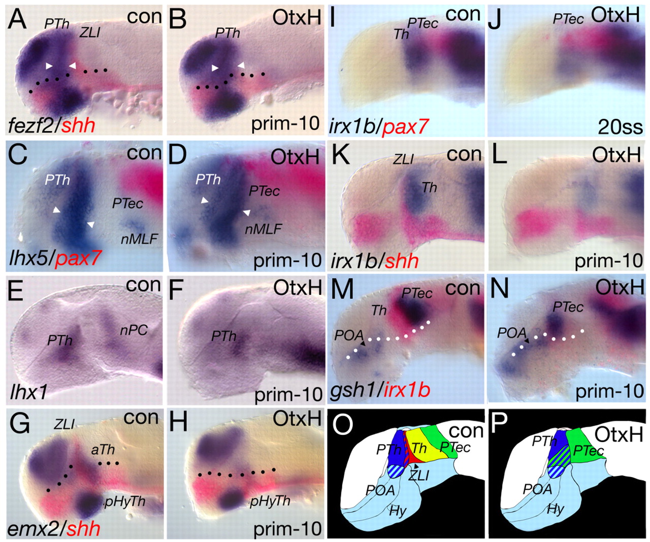
Fig. 4 Analysis of the MDT in OtxH embryos. (A-H) Single and double in situ hybridisation of OtxH embryos; the markers and stages are indicated. Shh expression is absent in the ZLI territory in OtxH embryos (B,H,L). Compared to control (con) siblings, markers for the prethalamic anlage - fezl, lhx5 and lhx1 - are still expressed in OtxH embryos and show a slight broadening in the ventral part of the anlage (lhx5: 11/15; fezl (fezf2): 12/20; A-F, marked by arrowheads). In OtxH embryos, the nuclei of the medio-longitudinal fascile is detectable by lhx5 (C,D), whereas the lhx1-positive nuclei of the PC are absent (E,F). emx2 expression in the anterior thalamus (aTh) is absent in OtxH embryos (23/30). (I-N) Prior to ZLI formation, at 20 somites (19 hpf), the irx1b expression domain is absent in OtxH embryos (I,J) and is still absent at prim-10 (28 hours; 24/60; K-N). (A-H) Dotted line marks the border between alar and basal plate. (C,D,M,N) pax7 is expressed in the dorso-lateral part of the pretectum (C,D), whereas gsh1 marks the ventro-medial domain of the pretectum (M,N). (C,D) In OtxH embryos, the pax7-positive part of the pretectum shifts anteriorly and a new border between the lhx5-positive prethalamus and the pax7-positive pretectum is observed (11/15). (M,N) Comparison of the gsh1-positive part of the pretectum in OtxH embryos and their wild-type siblings shows that the pretectum area abuts the gsh1-positive preoptic area (8/14). (O,P) Summary of the observations in OtxH embryos. In wild-type embryos, the MDT is organised in the following way: the lateral halves of the prethalamus (blue), the more medial ZLI (red), the thalamus (yellow) and the pretectum (green). In OtxH embryos, we still find the lateral halves of the prethalamus (blue) and the pretectum (green). Striped areas show the lateral to medial organisation of the area and not an overlap of expression domains. aTh, anterior thalamus; Hy, hypothalamus; nMLF, nucleus of the medio-longitudinal fascile; nPC, nucleus of the posterior commissure; pHyTh, posterior hypothalamus; POA, post-optic area; prim-10, primordium stage 10; PTec, pretectum; PTh, prethalamus; ss, somite stage; Th, thalamus; ZLI, zona limitans intrathalamica.

