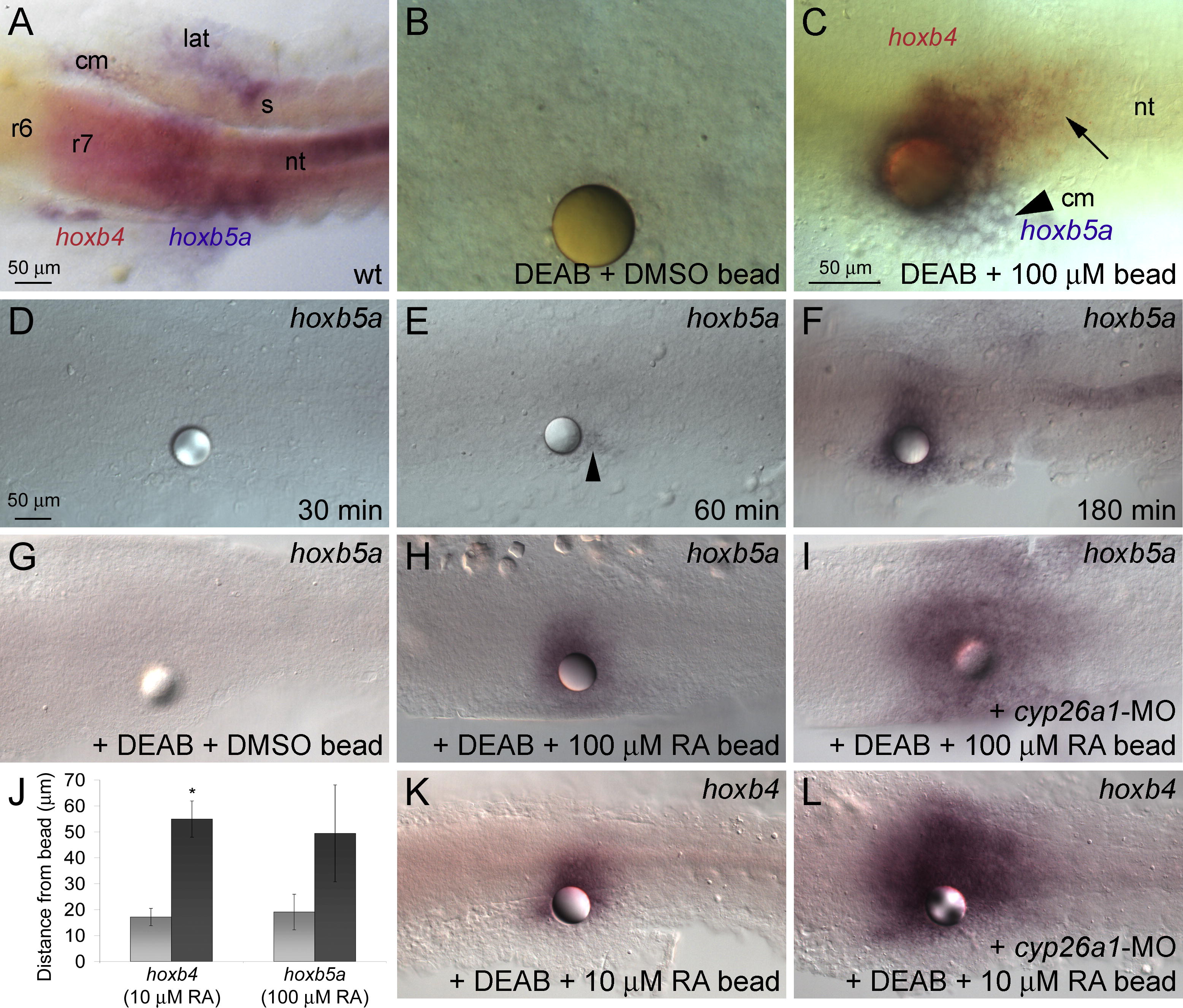
Fig. 2 Concentration-Dependent Induction of hox Gene Expression by RA (A) In wild-type (wt) embryos, hoxb4 expression in the neural tube (nt) extends to the r6/7 boundary, whereas the limit of hoxb5a expression is level with the first somite (s). hoxb5a is also expressed in axial, lateral (lat), and cranial (cm) mesoderm. (B and C) Beads soaked in 100 μM RA were implanted at 1?3 somites (10.3?11 hpf) into DEAB-treated embryos, fixed 3 h later, and assayed for hox expression. Dorsal views, showing in situ hybridization with probes for hoxb4 and hoxb5a at 14 hpf. (B) DMSO-coated beads do not induce hox expression. (C) RA-coated beads induce hoxb5a in lateral mesoderm (arrowhead), hoxb5a/hoxb4 double-positive cells near the bead, and hoxb4 single-positive cells further away (black arrow). (D?F) Time-course of hoxb5a induction in response to a 100 μM RA bead: no induction 30 min post-implantation (D), a few cells near the bead after 60 min (arrowhead) (E), and strong expression further away after 180 min (F). (G?L) RA-induced degradation limits the range of RA signaling. RA beads were implanted at 1?3 somites (10.3?11 hpf) into DEAB-treated embryos, fixed 3 h later, and assayed for hox expression. Dorsal views, showing in situ hybridization with probes for hoxb5a (G?I) or hoxb4 (K and L) at 14 hpf. (G) Implantation of a DMSO bead has no effect on gene expression in DEAB?treated embryos. (H and I) A 100 μM RA bead placed into a DEAB-treated, cyp26a1 morphant (I) induces hoxb5a over a much larger area than a DEAB-treated control (H). (J) Graph showing an increase in the range of hox induction in cyp26a1 morphant, DEAB-treated embryos (black bar) as compared to DEAB-treated (light grey bar). The asterisk (*) indicates significantly different from DEAB-treated controls (p < 0.05), using the Student t-test. (K and L) A 10 μM RA bead placed into a cyp26a1 morphant induces hoxb4 over a much larger area.
Image
Figure Caption
Acknowledgments
This image is the copyrighted work of the attributed author or publisher, and
ZFIN has permission only to display this image to its users.
Additional permissions should be obtained from the applicable author or publisher of the image.
Full text @ PLoS Biol.

