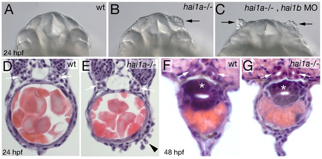
Fig. S1 Loss of Hai1 activity affects morphology of the olfactory epithelium, but not the morphology of the pronephric ducts and the gut. (A-C) Frontal views on head of live embryos at 24 hpf. Black arrows point to dissociating olfactory epithelium in hai1a mutant (with low penetrance and expressivity; B) and hai1a mutant injected with hai1b MO (with high penetrance and expressivity; C). (D-G) Eosin/hematoxylene stainings of cross-sections through posterior trunk region of hai1a mutants (E,G) and wild-type siblings (D,F) at 24 hpf (D,E) or 48 hpf (F,G). Pronephric ducts are indicated with white arrows, the gut in (F,G) with white asterisks. Black arrowhead in (E) points to dissociating epidermis on yolk extension.
Image
Figure Caption
Figure Data
Acknowledgments
This image is the copyrighted work of the attributed author or publisher, and
ZFIN has permission only to display this image to its users.
Additional permissions should be obtained from the applicable author or publisher of the image.
Full text @ Development

