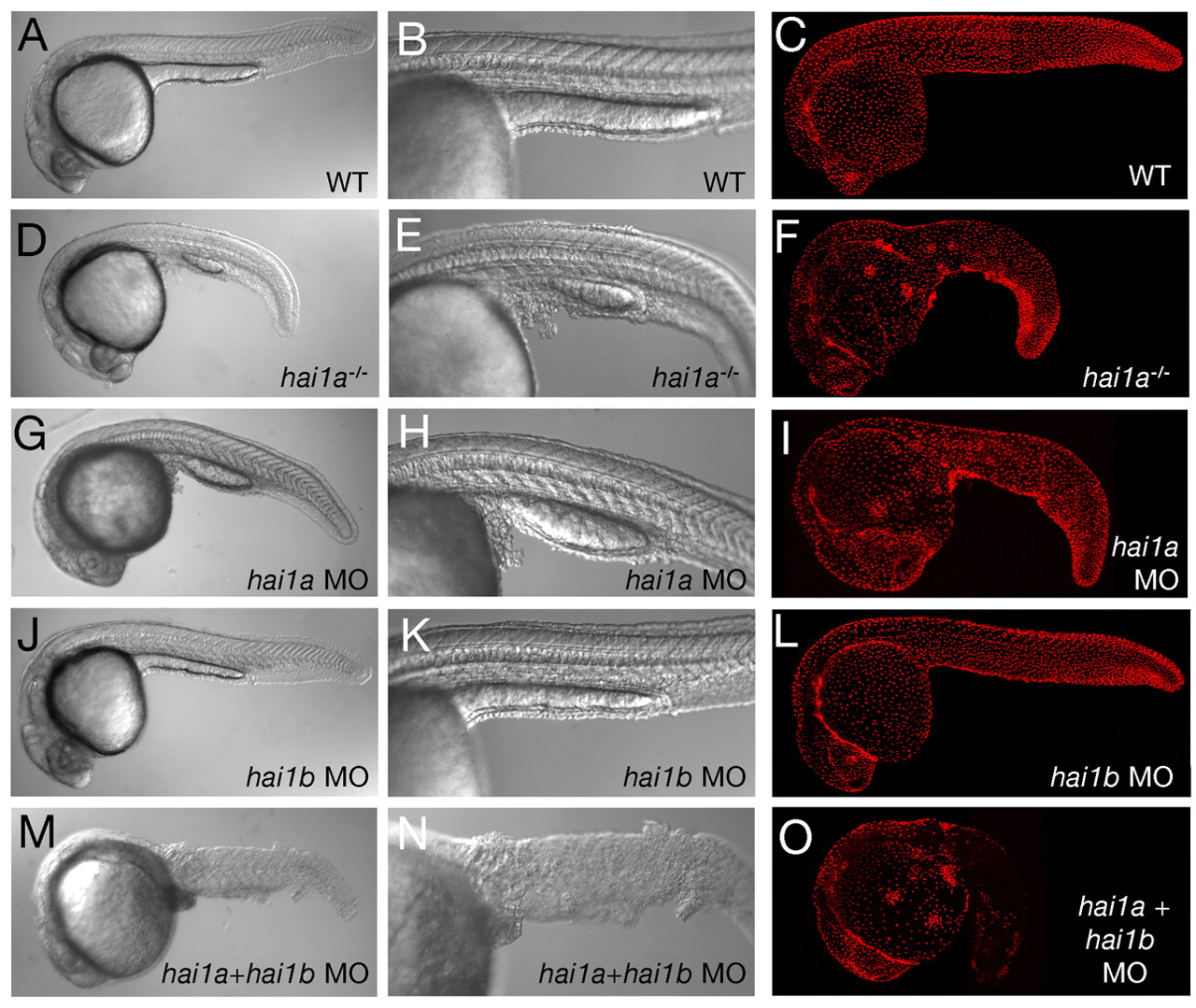
Fig. 3 Keratinocyte aggregation and shedding caused by the loss of Hai1a is further enhanced by the concomitant loss of its paralog, Hai1b. All panels show embryos at 24 hpf; wild-type siblings (WT; A-C), hai1a mutants (D-F), hai1a morphants (G-I), hai1b morphants (J-L) and hai1a mutants injected with hai1b MOs (M-O). Shown are lateral views of live embryos using Nomarski optics at low power (A,D,G,J,M) to assess overall embryo morphology, and at higher magnification (B,E,H,K,N) to assess epidermal defects in the trunk/tail regions, and lateral views of embryos after immunofluorescent detection of the basal epidermal marker protein p63 (C,F,I,L,O; merged stacks of confocal images).
Image
Figure Caption
Figure Data
Acknowledgments
This image is the copyrighted work of the attributed author or publisher, and
ZFIN has permission only to display this image to its users.
Additional permissions should be obtained from the applicable author or publisher of the image.
Full text @ Development

