
|
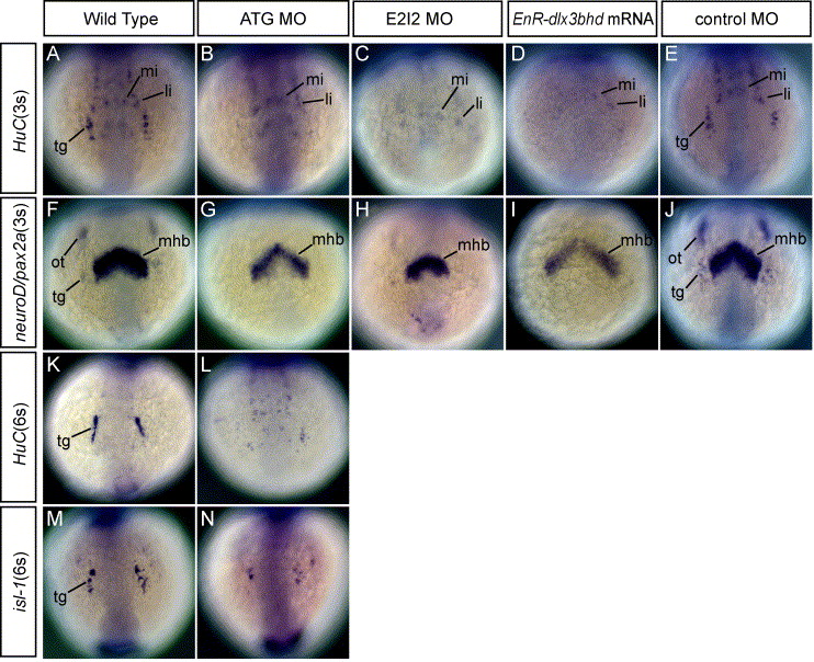
Fig. 3 Phenotypes of neurons in the trigeminal placodes in MOs and EnR-dlx3bhd mRNA injected embryos at 3- and 6-somite stage. (A–J) Embryos at 3-somite stage. Anterior views. (K–N) Embryos at 6-somite stage. Anterior views. (A, F, K, M) Wild-type embryos. (B, G, L, N) 20 ng ATG-dlx3b-MO/20 ng ATG-dlx4b-MO-injected embryos. (C and H) 20 ng E2I2-dlx3b-MO/20 ng E2I2-dlx4b-MO-injected embryos. (D and I) One hundred fifty-picogram EnR-dlx3bhd mRNA-injected embryos. (E and J) Forty-nanogram control MO-injected embryos. (A–E, K, L) Expression of HuC. (F–J) Expression of neuroD/pax2a. (M and N) Expression of isl-1. In a wild-type embryo (A), neurons in the trigeminal placodes (tg) and anterior lateral/medial hindbrain interneurons (li, mi) are observed. After injection of dlx3b and dlx4b-MOs or EnR-dlx3bhd mRNA shown in (B–D), anterior lateral/medial hindbrain interneurons remain, but neurons in the trigeminal placodes are highly reduced or absent as shown by expression of HuC. In a control-MO injected embryo (E), neurons in the trigeminal placodes are seen in the placodal primordial region. neuroD/pax2a expression in (G–I), both are highly reduced or absent in the placodal primordial region following injection of dlx3b and dlx4b-MOs or EnR-dlx3bhd mRNA. Midbrain–hindbrain boundary is seen in the central nervous system (mhb). In a control MO-injected embryos (J), no changes were observed in the trigeminal (tg) and otic placodal primordia (ot). Shown at the 6-somite stage, expression of HuC (L) and isl-1 (N), neurons in the trigeminal placodes remain highly reduced, as compared to wild-type embryos in (K and M), respectively.
Reprinted from Developmental Biology, 276(2), Kaji, T., and Artinger, K.B., dlx3b and dlx4b function in the development of Rohon-Beard sensory neurons and trigeminal placode in the zebrafish neurula, 523-540, Copyright (2004) with permission from Elsevier. Full text @ Dev. Biol.