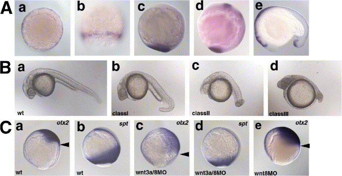
Fig. 1 Redundant role of Wnt3a and Wnt8 in early embryogenesis. (A) Expression of wnt3a. a, shield stage; b, 80% epiboly stage; c, bud stage; d, 8-somite stage (13 hpf); e, 17-somite stage (17 hpf). a, animal pole view with dorsal to the right; b, dorsal view; c, d, lateral view with dorsal to the right; e, lateral view with anterior to the left. (B) Phenotype of the wnt3aMO- and/or wnt8MO-injected pharyngula-stage (24 hpf) embryos. The phenotypes of the injected embryos were classified into three categories. Class I embryos had a ventrally bent tail structure and a normal number of somites (30?34). Class II embryos had an enlarged head, and a short and twisted tail (number of the somites 20?30; average 27.4 (n = 13)). Class III embryos had an enlarged head and a truncated tail structure (less than twenty somites; average 13.7 (n = 8)). The percentage of embryos in each class is shown in Table 1. (C) Expression of the anterior neuroectoderm marker otx2 (a, c, e) and the paraxial mesodermal marker spadetail (spt) (b, d) at the 90% epiboly stage in wild-type embryos (wt, a, b), and embryos injected with both wnt3aMO and wnt8MOs (wnt8-1MO and wnt8-2MO, c, d) or wnt8MOs alone (e). Lateral views with dorsal to the right. Arrowheads (a, c, e) indicate the posterior limits of the otx2 expression.
Reprinted from Developmental Biology, 279(1), Shimizu, T., Bae, Y.K., Muraoka, O., and Hibi, M., Interaction of Wnt and caudal-related genes in zebrafish posterior body formation, 125-141, Copyright (2005) with permission from Elsevier. Full text @ Dev. Biol.

