
|
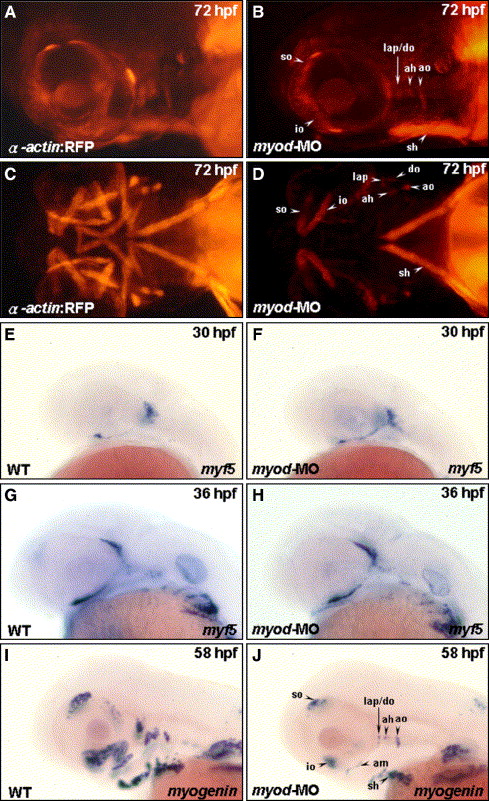
Fig. 4 Myod is required for the development of posterior extraocular recti and ventral branchial muscles. Embryos derived from the transgenic line Tg(α-actin:RFP) (A?D) and from the wild-type strain (E?J) were used. Embryos injected with 4 ng of myod-morpholino oligonucleotide (MO) to inhibit specifically myod translation were followed to observe the development of cranial muscle (B, D) and the expression of myf5 (F, H) and myogenin (J; lateral views: A, B, E?J; ventral views: C, D) at the stages indicated. The anterior extraocular recti (io and so), dorsal branchial muscle (ah, ao, do, and lap), and sh developed normally in the myod transgenic morphant (A vs. B; C vs. D). The posterior extraocular (sr, mr, ir, and lr) and the ventral branchial muscle (ima, imp, ih, and hh) were totally lost. When wild-type embryos were injected with myod-MO, myf5 was expressed normally in the myod morphants at 30 hpf (E vs. F) and at 36 hpf (G vs. H). Whereas, the expression of myogenin was decreased in myod morphants at 58 hpf (I vs. J), indicating a reduction in myogenin-positive muscle fibers.
Reprinted from Developmental Biology, 299(2), Lin, C.Y., Yung, R.F., Lee, H.C., Chen, W.T., Chen, Y.H., and Tsai, H.J., Myogenic regulatory factors Myf5 and Myod function distinctly during craniofacial myogenesis of zebrafish, 594-608, Copyright (2006) with permission from Elsevier. Full text @ Dev. Biol.