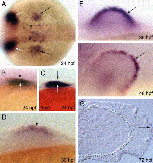
Fig. 1 Spatio-temporal profile of blimp-1 expression in the developing pectoral fin. (A) A wild-type embryo, showing blimp-1 expression in the fin bud (black arrows). Expression in the pharyngeal endoderm (white arrows) and spinal cord neurons (arrowheads) is also indicated. (B) Expression at this stage is evident in the ectoderm (black arrow) and the mesenchyme (white arrow). (C) tbx5 expression shows localization only to the mesenchyme (white arrow) and is excluded from the ectoderm (black arrow). (D) blimp-1 expression in the mesenchyme decreases and strengthens in the ectoderm (arrow). (E-G) blimp-1 expression in succeeding stages of embryogenesis. Panel A depicts dorsal view, all others depict lateral views. All panels of this and subsequent figures are oriented anterior to the left.
Reprinted from Developmental Biology, 300(2), Lee, B.C., and Roy, S., Blimp-1 is an essential component of the genetic program controlling development of the pectoral limb bud, 623-634, Copyright (2006) with permission from Elsevier. Full text @ Dev. Biol.

