
|
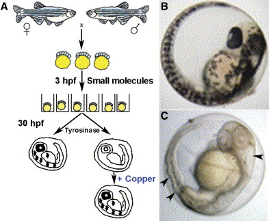
Fig. 1 Schematic of the small-molecule screen for drugs perturbing copper metabolism
A) Embryos from wild-type zebrafish were arrayed in a 96-well format and treated with drugs at 5 μM beginning at 3 hr postfertilization (hpf). At 30 hpf, the embryos were assayed for the development of melanin pigment. In a second screen, embryos were again exposed at 3 hpf to drugs that abrogated melanin formation but this time were supplemented with 25 μM CuCl2. In this screen, all drugs that prevented pigmentation in a manner reversible by the addition of exogenous copper were considered as perturbing copper homeostasis.
B and C) As compared to wild-type embryos (B), copper-deficient embryos treated with neocuproine (C) lacked pigment and displayed a strikingly wavy notochord and enlarged hindbrain ventricle (arrowheads).
Reprinted from Cell Metabolism, 4(2), Mendelsohn, B.A., Yin, C., Johnson, S.L., Wilm, T.P., Solnica-Krezel, L., and Gitlin, J.D., Atp7a determines a hierarchy of copper metabolism essential for notochord development, 155-162, Copyright (2006) with permission from Elsevier. Full text @ Cell Metab.