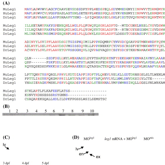
Fig. 3 Leg1 functions as a novel factor involved in controlling liver expansion growth. (A) Amino acid sequence alignment of Leg1 (DrLeg1, CT737258) with its homologues in human (HuLeg1, gi60502446) and mouse (MuLeg1) gi12844359). (B) RNA gel blot hybridization revealed that leg1 is undetectable before 17 hpf (lanes 1?5: 1-cell-stage, 5 hpf, 9 hpf, 12 hpf and 17 hpf, respectively). leg1 starts to express at 24 hpf and is maintained at relative high level through the rest of embryonic stages (lanes 6?10: 1 dpf, 2 dpf, 3 dpf, 4 dpf and 5 dpf, respectively). (C) Whole-mount in situ hybridization using a leg1 probe revealed that leg1 is enriched in the embryonic liver. (D) MOleg1 morphants conferred a smaller liver phenotype (35 out of 42 morphants examined; representative embryo shown on the left) when compared with that of a control MOmis morphant (embryo on the right). Co-injection of the leg1 mRNA with MOleg1 restored the morphant phenotype to normal (embryo in the middle; 26 out 38 embryos examined). Embryos were stained with triple markers: a secreted immunoglobulin 4 probe as the liver (lv) marker, an insulin probe as the endocrine pancreas (en) marker and a trypsin probe as the exocrine pancreas (ex) marker.
Reprinted from Developmental Biology, 294(2), Cheng, W., Guo, L., Zhang, Z., Soo, H.M., Wen, C., Wu, W., and Peng, J., HNF factors form a network to regulate liver-enriched genes in zebrafish, 482-496, Copyright (2006) with permission from Elsevier. Full text @ Dev. Biol.

