
Image
|
Figure Caption
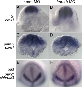
Fig. 3 Neural tube closure and dorsoventral patterning are normal in lmo4b morphant embryos. Panels A?D are cross sections through the anterior neural rod (A, B) or neural tube (C, D) in the region of the forebrain from control (A, C) and morphant (B, D) embryos. In situ hybridization probes used to localize forebrain tissue indicated at the left. (E, F) Bud stage embryos stained with probes to indicate the size of the anterior neural plate.
Figure Data
Acknowledgments
This image is the copyrighted work of the attributed author or publisher, and
ZFIN has permission only to display this image to its users.
Additional permissions should be obtained from the applicable author or publisher of the image.
Reprinted from Developmental Biology, 309(2), McCollum, C.W., Amin, S.R., Pauerstein, P., and Lane, M.E., A zebrafish LMO4 ortholog limits the size of the forebrain and eyes through negative regulation of six3b and rx3, 373-385, Copyright (2007) with permission from Elsevier. Full text @ Dev. Biol.