Fig. 2
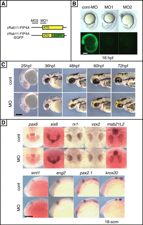
Fig. 2 Small eye and head phenotype in zRab11-FIP4-depleted embryos. (A) Schematic representation of the structure of zRab11-FIP4A-EGFP fusion gene and corresponding positions of the zRab11-FIP4A-specific MOs. Colored bars and solid lines indicate ORFs and untranslated regions, respectively. (B) Evaluation of the effect of the zRab11-FIP4A-specific MOs. Embryos were injected with in vitro synthesized zRab11-FIP4A-EGFP RNA along with control-MO, zRab11-FIP4-MO1 or MO2. Morphology (upper) and EGFP expression (lower) at 18 hpf are shown in lateral views. (C) Morphology of control embryos (upper) and zRab11-FIP4 morphants (lower) observed at the indicated times is shown in lateral views. (D) Expression of retinal and brain markers in an early stage of MO-injected embryos. Control-MO- or zRab11-FIP4-MO1-injected embryos were fixed at 18 hpf and then subjected to whole-mount in situ hybridization. In all cases, expression of markers in zRab11-FIP4 morphants was comparable to that in control embryos. Scale bars indicate 200 μm.
Reprinted from Developmental Biology, 292(1), Muto, A., Arai, K.I., and Watanabe, S., Rab11-FIP4 is predominantly expressed in neural tissues and involved in proliferation as well as in differentiation during zebrafish retinal development, 90-102, Copyright (2006) with permission from Elsevier. Full text @ Dev. Biol.

