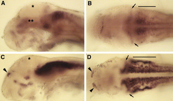
Fig. 6 RNA expression patterns of robo3 isoforms during the second day of development. Lateral (A, C) and dorsal (B, D) views of the head showing whole-mount RNA in situ hybridizations for robo3var1 and robo3var2 at 48 hpf. (A, B) Diffuse expression of robo3var1 in the midbrain (double asterisks in A) with stronger expression in the cerebellum (B, arrows) and hindbrain (B, bar). (C, D) Distinct and robust expression of robo3var2 in the cerebellum (D, arrows) and hindbrain (D, bar). Notice the lack of expression of both isoforms in the tectum (A and C, asterisks). Patches of robo3var2 expressing cells are present in the diencephalic regions (C, arrowhead) and in the telencephalon (D, arrowheads).
Reprinted from Mechanisms of Development, 122(10), Challa, A.K., McWhorter, M.L., Wang, C., Seeger, M.A., and Beattie, C.E., Robo3 isoforms have distinct roles during zebrafish development, 1073-1086, Copyright (2005) with permission from Elsevier. Full text @ Mech. Dev.

