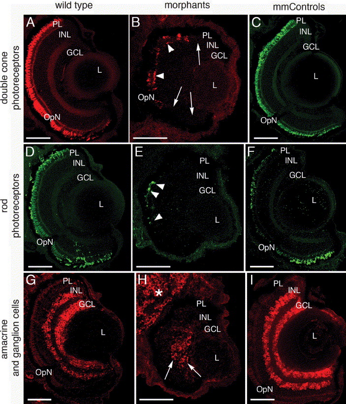
Fig. 7 Reduction in retinal neurons revealed by immunohistochemistry at 5 dpf. Wild type (left column), morphant (center column) and mismatch control (right column) retinal cell types were identified by immunolocalization of zpr-1 (double cone photoreceptor cells, A–C), rhodopsin (rod photoreceptor outer segments, D–F), and HuC/D (amacrine and ganglion cells, G–I) in frozen eye sections. While some double cone cells are identified in the morphant (B, arrowheads), large regions of the retina lack this cell type (B, arrows). Similarly, only small numbers of rhodopsin-positive photoreceptors were detected in the morphants (E, arrowheads) compared to the wild type (D) and mismatch control retinas (F). The amacrine and ganglion cells (G–I) were also reduced in the morphants and were characterized by an absence of immunolabeling towards the retinal margins (compare H to G and I). Abbreviations: PL, photoreceptor layer; INL, inner nuclear layer; GCL, ganglion cell layer; OpN, optic nerve; L, lens. The scale bars represent 50 μm.
Reprinted from Mechanisms of Development, 122(4), Shi, X., Bosenko, D.V., Zinkevich, N.S., Foley, S., Hyde, D.R., Semina, E.V., and Vihtelic, T.S., Zebrafish pitx3 is necessary for normal lens and retinal development, 513-527, Copyright (2005) with permission from Elsevier. Full text @ Mech. Dev.

