
Fig. 2
|
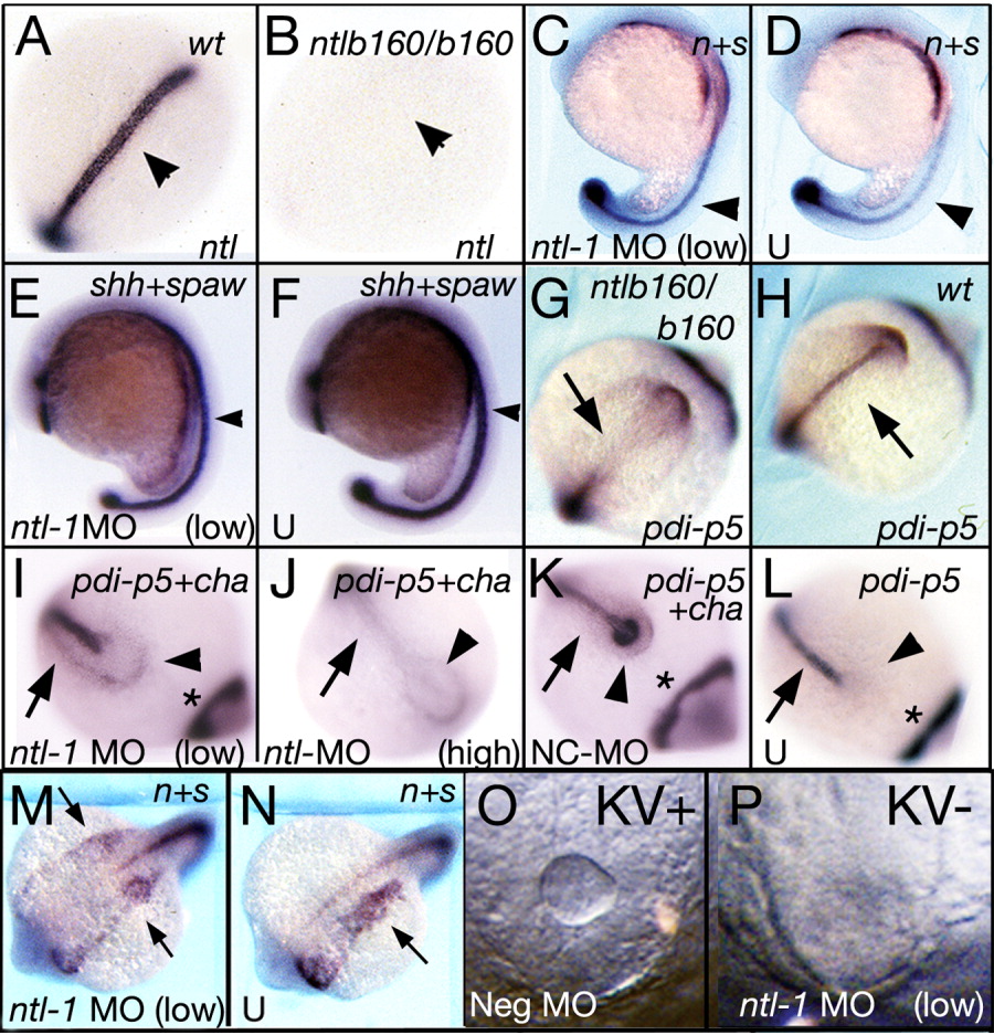
Fig. 2 Characterization of low-dose ntl morphant embryos. A,B: Seven to 12 s, dorsal view. From a ntlb160 incross. Arrowhead: notochord. A: Wild-type sib. B: ntlb160 homozygote. C,D: Sixteen to 19 s, lateral view. n+s: ntl + spaw probes. Arrowhead: ntl mRNA in the notochord. C: Injected with 0.3 ng ntl-1 MO. D: U: Uninjected. E,F: Sixteen to 19 s, lateral view. sonic hedgehog (shh; arrowhead) + spaw probes. E: Injected with 0.2 ng ntl-1 MO. F: U: Uninjected. G-L: Arrows: Site of pdi-p5 expression in posterior notochord. Arrowhead: site where cha is normally expressed. G,H: Ten to 14 s. Dorsal view, close-up of tail and tailbud. Head to upper right. Two embryos from a ntlb160 incross. pdi-p5 probe. I-L: Dorsal view of tailbud/KV region, head to lower right. pdi-p5� cha probes. Asterisk: pdi-p5 staining in anterior (out of field of view in J). I-K: Nine to 12 s. L: Seven to 9 s. I: Injected with 0.1 ng ntl-1 MO. J: Injected with 4 ng of ntl-1 MO. K: Injected with 4 ng NC-MO. L: U: Uninjected. M,N: Sixteen to 19 s, dorsal views of the same embryos shown in Figure 2C and D, respectively. Head at lower left, ntl + spaw probes. Arrows: spaw mRNA in lateral plate. M: Injected with 0.3 ng ntl-1 MO. N: U: uninjected. O,P: Nine to 12 s. Brightfield view of tailbud/KV region, live embryos. KV� denotes presence or absence of a visible Kupffer's Vesicle. Neg. MO: scrambled antisense cha MO. O: Injected with 0.85 ng Neg. MO. P: Injected with 0.5 ng ntl-1 MO.