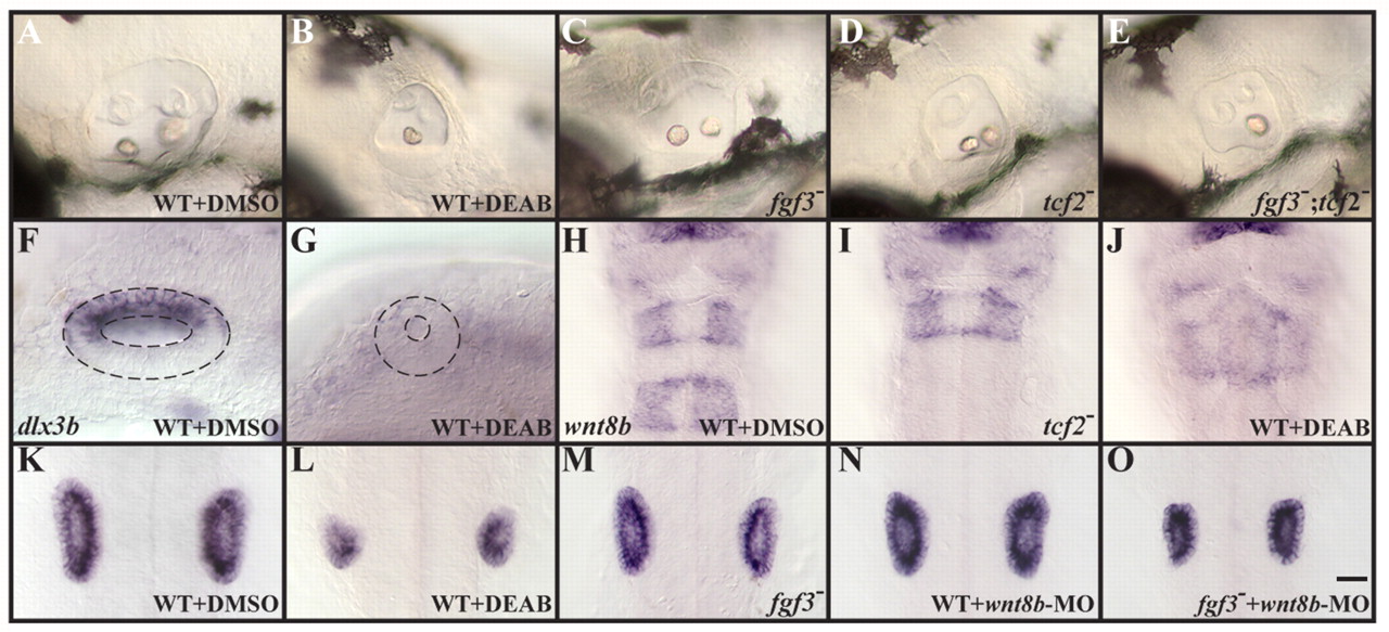
Fig. 7 Rhombomere 5-specific Wnt8b function is required for the maintenance of otic fate. (A-D) Assessed by morphology, otic vesicles are reduced in fgf3 (C) and tcf2 (D) single mutants compared to wild-type embryos (A), but not as much as in RA-depleted embryos (B). (E) Loss of both fgf3 and tcf2 together results in otic vesicles that form only one otolith, as is observed in RA-depleted embryos (B). (F,G) Expression of dlx3b in the dorsal half of the otocyst at 22 hpf is lost in embryos depleted of RA signaling. (H-J) In control embryos at 22 hpf, wnt8b expression can be detected in the midbrain-hindbrain boundary, in r3 and in r5 (H), whereas tcf2 mutants (I) and RA-depleted embryos (J) show wnt8b in the midbrain-hindbrain boundary and in r3, but not in r5. (K-N) Inactivation of Wnt8b in wild-type embryos by morpholino injection (MO, N) leads to a reduced ear size (labeled with stm at 22 hpf), similar to the size observed in fgf3 mutants (M), compared to untreated wild type (K); however, the size reduction is not as severe as in embryos depleted of RA signaling (L). Loss of Wnt8b function in fgf3 mutants (O) produces reduced otic vesicles comparable to RA-depleted embryos (L). (A-G) Lateral views with anterior to the left and dorsal towards the top. (H-O) Dorsal views with anterior towards the top. Scale bar: 35 µm for A-E and H-O; 20 µm for F,G.
Image
Figure Caption
Figure Data
Acknowledgments
This image is the copyrighted work of the attributed author or publisher, and
ZFIN has permission only to display this image to its users.
Additional permissions should be obtained from the applicable author or publisher of the image.
Full text @ Development

