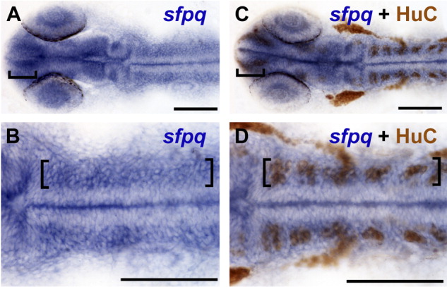
Fig. 4 sfpq is strongly expressed in regions of neurogenic activity. A-D: At 24 hours postfertilization (hpf) wild-type embryos labeled for sfpq expression by in situ hybridization only (A,B) and wild-type sibling embryos double labeled for sfpq mRNA expression and HuC protein by immunohistochemistry (C,D) show that regions of strongest sfpq expression in the brain (brackets) overlap with HuC, a marker for postmitotic neuronal precursors. B,D: Higher magnification of hindbrain; brackets mark strong sfpq expression overlapping with HuC labeling. Midline staining is an artifact of the staining process, because it is not observed in embryos cut open before staining. A-D: Dorsal views. Anterior left. Scale bar = 100 μm.
Image
Figure Caption
Figure Data
Acknowledgments
This image is the copyrighted work of the attributed author or publisher, and
ZFIN has permission only to display this image to its users.
Additional permissions should be obtained from the applicable author or publisher of the image.
Full text @ Dev. Dyn.

