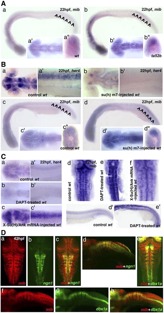
Fig. 6 mib expression is negatively regulated by Su(H)-dependent Notch activation. (A) mib expression in (a) wt and (b) mibta52b embryos at 22hpf, lateral view. The inserts show the mib expression in (a′?b′) the brain region, dorsal view and (a″?b″) a transverse section across posterior trunk. (B) her4 expression in (a?a′) control wt and (b?b′) su(h)-m7-injected embryos at 22hpf; (a?b) head region, dorsal view; (a′?b′) trunk region, lateral view. mib expression in (c) control wt and (d) su(h)-m7-injected embryos at 22hpf, lateral view. The inserts show the mib expression in the (c′?d′) head region, dorsal view and (c″?d″) a transverse section across posterior trunk. (C) her4 expression in (a?a′) control wt, (b?b′) DAPT-treated and (c?c′) X-Su(H)/Ank mRNA-injected embryos at 22hpf; (a?c) head region, dorsal view; (a′?c′) trunk region, lateral view. mib expression in (d?d′) control wt, (e?e′) DAPT-treated, and (f) X-Su(H)/Ank mRNA-injected embryos at 22hpf; (d?f) mid- and hindbrain region, dorsal view; (d′?e′) trunk and tail region, lateral view. (D) Coexpression of mib with ngn1 or dbx1a in hindbrain differentiating neurons at 42hpf. (a?c) and (e) are dorsal view; (d) and (f?h) are lateral view. Note that ngn1 and dbx1a are coexpressed with mib in different subpopulations of neurons.
Reprinted from Developmental Biology, 305(1), Zhang, C., Li, Q., Lim, C.H., Qiu, X., and Jiang, Y.J., The characterization of zebrafish antimorphic mib alleles reveals that Mib and Mind bomb-2 (Mib2) function redundantly, 14-27, Copyright (2007) with permission from Elsevier. Full text @ Dev. Biol.

