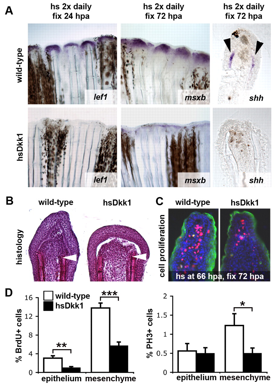
Fig. 3 Wnt/β-catenin signaling regulates specification and proliferation of the regeneration blastema. (A) Expression of lef1, a marker for the basal epidermal layer of the regeneration epithelium, msxb, marking the mesenchymal progenitor cells of the blastema, and shh, expressed in basal epidermal cells (shown in thick sections), is strongly reduced in Dkk1-overexpressing fins. lef1 is shown at 24 hpa (n=4), msxb (n=4) and shh (n=4) at 72 hpa. Fish were heat shocked twice daily starting shortly before amputation. (B) Hematoxylin-stained sections of tail fin regenerates at 48 hpa. Dkk1-overexpressing fins (right panel; n=6) display reduced numbers of deep mesenchymal cells of the blastema. Fish were heat shocked twice daily starting shortly before amputation. Arrowheads indicate the plane of amputation. (C) 72 hpa regenerates stained for BrdU (red), phosphorylated histone H3 (PH3, green) and DAPI (blue). Cell proliferation in both the mesenchyme and epithelium is decreased in Dkk1-overexpressing fins. Fish were heat shocked once at 66 hpa and fixed at 72 hpa. (D) Quantification of the cell proliferation defects in Dkk1-overexpressing regenerating fins. The fraction of BrdU-positive (left) and PH3-positive (right) cells relative to the total number of cells (DAPI-positive) is shown in percent (n=11). Error bars represent the s.e.m; *P=0.0495; **P=0.0025; ***P=7.076x10⁻⁶ (two-tailed).
Image
Figure Caption
Figure Data
Acknowledgments
This image is the copyrighted work of the attributed author or publisher, and
ZFIN has permission only to display this image to its users.
Additional permissions should be obtained from the applicable author or publisher of the image.
Full text @ Development

