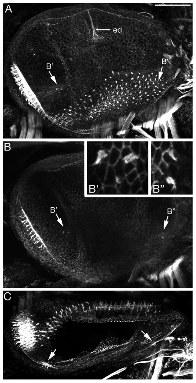
Fig. 4 Morphology of the L. fluviatilis otic vesicle at stage 30. (A-C) Projections from confocal z-stacks of a FITC-phalloidin-stained ear showing the actin-rich sensory hair cell stereociliary bundles and general morphology. (A,B) Lateral views, anterior to left, dorsal to top; both are composites of two images. ed, endolymphatic duct. (B) A projection from a lateral subset of the confocal z-stack to show the presumptive cristae clearly, also shown at higher power in insets (B',B''). (C) Dorsal view, anterior to left, medial to top; arrows indicate hair cells in the developing cristae. Scale bars: 50 μm.
Image
Figure Caption
Acknowledgments
This image is the copyrighted work of the attributed author or publisher, and
ZFIN has permission only to display this image to its users.
Additional permissions should be obtained from the applicable author or publisher of the image.
Full text @ Development

