
Image
|
Figure Caption
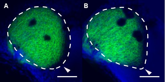
Fig. S3 The Tectum of exa Mutants Has an Abnormal Shape RGC axon tracing, following whole-eye DiI fills at 7 dpf, reveals a subtle extension of the tectal neuropil (delineated by DAPI counterstaining) at the ventral-posterior margin (arrow). Scale bar is 50 μm
Figure Data
Acknowledgments
This image is the copyrighted work of the attributed author or publisher, and
ZFIN has permission only to display this image to its users.
Additional permissions should be obtained from the applicable author or publisher of the image.
Full text @ PLoS Genet.