
Image
|
Figure Caption
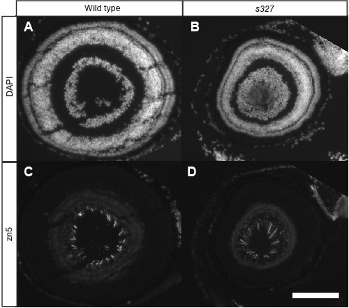
Fig. S2 Dorsal RGCs Are Present and Properly Differentiated in darl Mutants Sagittal sections of WT (A and C) and darls327 retina (B and D) were stained with DAPI (A and B) and zn5 (C and D), a marker for differentiated RGCs. RGCs are present in the dorsal part of the retina and sending out axons into the optic nerve head in the mutant. The mutant eyes are reduced in size compared to WT.
Figure Data
Acknowledgments
This image is the copyrighted work of the attributed author or publisher, and
ZFIN has permission only to display this image to its users.
Additional permissions should be obtained from the applicable author or publisher of the image.
Full text @ PLoS Genet.