
|
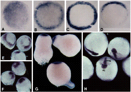
Fig. 8 Spatial localization of goosecoid transcripts in lithiumized embryos. Stages are +/- 15 minutes. Embryos in A-G and in H were exposed to 0.3 M LiCl for 10 minutes at 2 h and 4 h, respectively. For A-D, the yolks were removed and the blastoderms were photographed in bright field. (A) Animal pole view of a 4 h embryo. Staining is observed throughout the blastoderm; this staining is typically slightly more intense in a portion of the blastoderm. (B) Animal pole view of a 5 h embryo. While significant hybridization still occurs throughout the blastoderm, the staining is largely concentrated around the margin. (C) Animal pole view of 6 h embryo, radialized shield. Cells around the entire circumference of the germ ring are stained. (D) Animal pole view of a 6 h embryo, broad shield. Staining is restricted to a 160° arc at the margin. Note that lateral observation of the 6 h embryos shows that staining is restricted to involuted cells. For E-H, the yolks were left intact, and the embryos were photographed in dark field. (E) Oblique animal pole view of 8 h embryos. A ring of hybridization is observed above the germ-ring/yolk margin. (F) Oblique animal pole view of 10 h embryos. Hybridization is just ventral to the animal pole. (G) Lateral view of 14 h embryos. A circular cap of staining occupies the animal pole. (H) Dorsal and anterior views of 10 h late-lithium embryos. While the medial-strip (ms) and medial swelling of the rostral-crescent (rc) patterns are observed, the lateral-wing staining is reduced or absent.