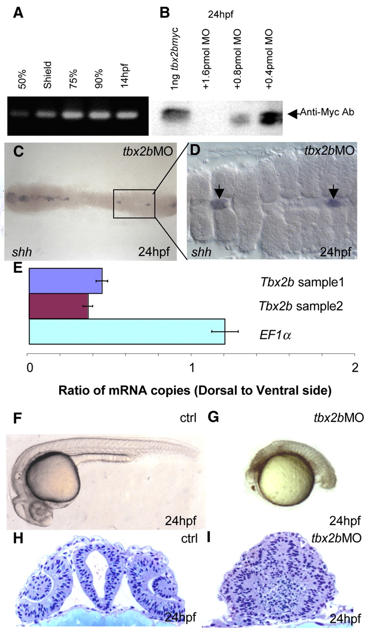
Fig. 1 tbx2b MO blocks translation of tbx2b and impairs normal development. (A) RT-PCR using primers specific to the 5'-UTR of tbx2b shows that this transcript was present as early as 50% epiboly. (B) Western blot with anti-Myc antibody shows that translation of recombinant tbx2b-myc was blocked in the presence of tbx2b MO in a dose-dependent manner. (C) Dorsal view of tbx2b morphant shows the almost complete loss of shh in the trunk region of the embryo. The boxed section is enlarged in D, showing the shh-positive cells as remnants of notochord (arrow). (E) Real-time PCR shows that tbx2b transcripts are present at a higher level in the ventral gastrula (5.5 hpf) when compared with EF1α. (F,G) Phenotype of tbx2b morphant (2 pmol) (G) when compared with control (F) (see text for description). (H,I) tbx2b is required for proper development of the eyes and forebrain. (H) Plastic cross-section through the forebrain of a 24 hpf control embryo at the level of the lens. (I) Cross-section of the tbx2b morphant shows severe disorganization of the forebrain and eyes.
Image
Figure Caption
Figure Data
Acknowledgments
This image is the copyrighted work of the attributed author or publisher, and
ZFIN has permission only to display this image to its users.
Additional permissions should be obtained from the applicable author or publisher of the image.
Full text @ Development

