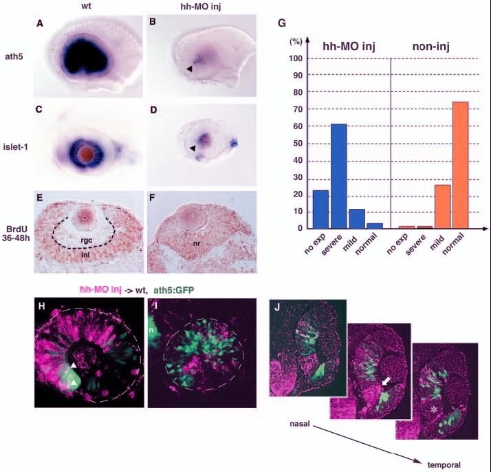
Fig. 7 Shh and Twhh regulate the progression of ath5 expression. (A,B) ath5 expression in 33-hpf wild-type (A) and Hh-MO-injected (B) retinas. ath5 expression fails to progress from the ventronasal retina injected with Hh-MO (arrowhead). (C,D) islet1 expression in 48-hpf wild-type (C) and Hh-MO-injected (D) retinas. islet1 expression fails to progress from the ventronasal retina injected with Hh-MO (arrowhead). (E,F) Labelling of 48-hpf wild-type (E) and Hh-MO injected (F) retinas with an anti-BrdU antibody (brown). Because BrdU is incorporated from 36 to 48 hpf, the RGC layer is BrdU negative in the wild-type retina. By contrast, almost all retinal cells are labelled with the anti-BrdU antibody in the Hh-MO-injected retina. (G) Quantitative assessment of ath5 expression in Hh-MO-injected (blue) and non-injected control (orange) embryos. The defect in ath5 expression in Hh-MO-injected embryos shows a similar profile to that in Gli-MO-injected embryos (blue bars in Fig. 4M). Numbers of Hh-MO-injected and non-injected embryos examined were 33 and 11, respectively. (H) Hh-MO-injected cells were incorporated into 36-hpf wild-type peripheral retina. ath5:GFP expression (green) does not progress towards the Hh-MO-positive area (magenta), although double-positive cells (white arrowheads) are observed at the interface between wild-type and Hh-MO areas. (I) Hh-MO-injected cells were incorporated into 36 hpf wild-type central retina. Hh-MO (magenta) and ath5:GFP (green) are segregated. (J) Three adjacent sections are shown along the axis from the nasal to temporal regions. ath5:GFP expression (green) does not occur in retinal columns derived from Hh-MO-injected donor embryos (magenta). GFP is not expressed even in wild-type retinal columns (white asterisk in right panel) located adjacent to the temporal side of Hh-MO-derived retinal columns (white arrow in middle panel). inl, inner nuclear layer; n, nose; nr, neural retina; rgc, retinal ganglion cell layer.
Image
Figure Caption
Figure Data
Acknowledgments
This image is the copyrighted work of the attributed author or publisher, and
ZFIN has permission only to display this image to its users.
Additional permissions should be obtained from the applicable author or publisher of the image.
Full text @ Development

